Download
1 / 72
810 likes | 1.19k Views
REPRODUCTIVE ANATOMY & PHYSIOLOGY. EARLY DEVELOPMENT. Male & Female organs produce sex cells transport for union Sex Differentiation at 8 weeks of life Ovary - produces oogonia at 10 weeks of fetal life; approximately 150,000 oocytes present at birth

E N D
EARLY DEVELOPMENT • Male & Female organs • produce sex cells • transport for union • Sex Differentiation at 8 weeks of life • Ovary - produces oogonia at 10 weeks of fetal life; approximately 150,000 oocytes present at birth • Testes - produces spermatoza at 7-8 weeks
Reproductive Anatomy • External Organs • Mons Pubis • Labia Majora • Labia Minora • Clitoris • Vaginal Vestibule • Urethral meatus • Skene’s Glands • Hymen • Fourchete • Perineum
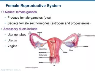
Reproductive Anatomy con. • Internal Organs • Vagina • Uterus • Fundus • Corpus • Isthmus • Cervix
Layers of the Uterus • Perimetrium • outer layer composed of peritoneum • Myometrium • inner layer primarily in the fundus; longitudinal fibers; causes cervical effacement and power to express the baby • Endometrium • innermost layer, produces endometrial milk, undergoes monthly regeneration
Myometrium • Muscular Layer - composed of 3 distinct layers • Longitudinal fibers found mainly over the fundus; most involved with birth of fetus • Fibers interlaced with blood vessels in Figure 8 pattern; living ligature – helps stop bleeding • Circular fibers concentrated around fallopian tubes and cervical os; helps keep cervix closed
Internal Organs con. • Isthmus • Joins corpus to the cervix • Site for lower C/S • Cervix • Composed of fibrous connective tissue • Length 2.5 to 3 cm (~1-2”) • Functions • Passage of menses and sperm • Produces mucus in response to cyclic hormones • Frequent site for uterine cancer
Uterine LigamentsThink which ligaments cause pain during pregnancy • Broad ligament – stabilizes uterus, covers uterus anteriorly and posteriorly • Round ligament – helps keep uterus in place from the sides, pain on sides late in pregnancy • Ovarian ligament – anchors lower part of ovary, helps catch ovum in fimbriae • Cardinal ligament – chief uterine support, prevents uterine collapse • Uterosacral ligament – support for uterus at level of the ischial spine, source of menstrual pain
Fallopian Tubes • Functions – provide passageway for ovum into uterus, site for fertilization • Fimbriae – most distal part, wavelike motion that pulls ovum into tube • Ampulla – site for fertilization • Isthmus - close to uterus, site for BTL
Ovaries • At birth, all ova are contained within immature follicles • Functions • Ovulation • Produce hormones
Bony Pelvis • Functions – to support and protect the internal organs of reproduction • Innominate Bones • Ilium – upper prominence of hip • Ischium – under the ilium, ends in ischial tuberosity, serves as reference point for station • Pubis – (2 separate bones) front of innominate, meets other to form symphysis pubis • Sacrum – 5 fused vertebrae, sacral promontory • Coccyx – (Tail bone) triangular bone last on vertebral column, moves backward in childbirth (Sometimes can get fx’d during childbirth)
Pelvic Floor (Muscles) • Designed to overcome force of gravity • Provides stability and support for surrounding structures (Help body remain intact, until baby is ready for birth) • Pelvic diaphragm – deep fascia, levator ani, and coccygeal muscles • Muscles function as a whole, yet are able to move over one another – provides capacity for dilatation
Figure 2–9 Muscles of the pelvic floor. (The puborectalis, pubovaginalis, and coccygeal muscles cannot be seen from this view.)
Pelvic Division • False Pelvis – portion above pelvic brim or inlet; serves to support pregnant uterus; helps direct presenting part into true pelvis • True Pelvis – portion below linea terminalis; represents the bony limits of the birth canal • Pelvic inlet – upper border of true pelvis • Pelvic outlet – lower border of true pelvis
Figure 2–10a Female pelvis. False pelvis is shallow cavity above the inlet; true pelvis is deeper portion of cavity below the inlet.
Figure 2–10b True pelvis consists of inlet, cavity (midpelvis), and outlet.
Figure 8–5a Manual measurement of inlet and outlet. Estimation of the diagonal conjugate, which extends from the lower border of the symphysis pubis to the sacral promontory.
Figure 8–5b Estimation of the anteroposterior diameter of the outlet, which extends from the lower border of the symphysis pubis to the tip of the sacrum.
Pelvic MeasurementsHelps figure whether baby’s head can fit. • Diagonal conjugate – extends from the subpubic angle to the middle of the sacral promontory; can be measured manually (with hand)during a pelvic exam • Take and substract 1.5cm to get Obstetric conjugate. • Obstetric conjugate – extends from the middle of the sacral promontory to 1 cm below the pubic crest (Cannot be reached/measured manually) • Conjugate vera – extends from the middle of the sacral promontory to the pubic crest
Figure 2–11 Pelvic planes: coronal section and diameters of the bony pelvis.
C D Figure 8–5 c & d Methods that may be used to check the manual estimation of anteroposterior measurements.
Figure 8–6 Use of a closed fist to measure the outlet. Most examiners know the distance between their first and last proximal knuckles. If they do not, they can use a measuring device.
Pelvic Types • Gynecoid – most common female, adequate • Android – most common male, not adequate • Anthropoid – usually adequate • Platypelloid – usually not adequate
Female Sex Hormones • Estrogen • Maturation of secondary sex characteristics • Secreted by the maturation of ovarian follicles • Cause proliferation of endometrial mucosa • Causes increase in size and weight; closure of long bones • Increases myometrial and fallopian tube contractility • Increases uterine sensitivity to oxytocin • Maintains bone density • Inhibits FSH production and stimulates LH production • May increase libido
Female Sex Hormones con. • Progesterone “keeps everything quiet”; maintains pregnancy • LH stimulates corpus luteum to secrete progesterone • Decreases motility and contractility of uterus • Proliferates vaginal epithelium • Causes cervix to secrete thick viscous mucus • Anti-sperm • Prepares breast tissue for lactation • Thermogenic “heat producing” • check temp to determine ovulation • “Hormone of Pregnancy”
Female Sex Hormones con. • Prostaglandins • Produces by the endometrium “lining of the uterus” • Differentiated by Roman letters and numbers or Greek numbers • Essential for ovulation (help egg be expelled from the ovary) • Causes expulsion of the ovum • Produces progesterone withdrawal • Facilitates tissue digestion to cause endometrial shedding
Neurohormonal Basis of the Female Reproductive CycleCauses menses cycle to occur • Controlled by an interaction between the nervous and endocrine systems and their target tissues – hypothalamus, anterior pituitary, and ovaries
NEUROHORMONAL BASIS OF THE FRC • CNS --- HYPOTHALMUS RELEASES ----GnRF (FSHRH & LHRH) ------CAUSES ANTERIOR PITUITARY TO RELEASE FSH & LH ---- STIMULATES GONADS TO SECRETE HORMONES (ESTROGEN & PROGESTERONE) • In males, LH induces secretion of testosterone. • In females, LH working w/ FSH stimulate follicle growth in ovary to secrete estrogen.
Ovarian CycleBe able to know which cycle the woman is in • 1st Part- Follicular Phase (Follicles- where the immature eggs are contained) • Starts with day 1 menses; 1-14 days; may vary • multiple follicles are maturing; one is selected; when mature, called a graafian follicle; surrounded by fluid …and becomes a cyst on the ovary. • Comes close to surface of ovary, forms a blister, ovum pushed out of the follicle near the fimbria (ovulation) • Pain at mid-cycle Mittelschmerz (may see blood spotting) • Pulled into fallopian tube and travels to ampulla where fertilization can occur
Ovarian Cycle con. • 2nd Part- Luteal Phase • Begins with ovulation (the second half of ovarian cycle) • Corpus luteum develops from the ruptured follicle • If fertilized, the ovum implants into endometrium • Ovum secretes HCG to maintain corpus luteum; the corpus luteum secretes progesterone and estrogen (cause you have to have high levels of H to maintain preg) • If no fertilization, degenerates in about a week and becomes the corpus albicans • Estrogen and Progesterone decrease which stimulates FSH and LH to be released which will start whole cycle over again. • 14 days after ovulation, menses begins (this remains constant) A person can then predict ovulation.
Figure 2–14 Various stages of development of the ovarian follicles.
Endometrial Cycle- 4 Phases • 1) Menstruation • Cyclic uterine bleeding in response to hormones changes; begins 14 days after ovulation • Partial shedding of the endometrium • Discharge made up of blood, fluid, cervical and vaginal secretions, bacteria, leukocytes and cellular debris; dark red, distinctive odor • Menarche- onset of menstruation; age 9-16 • Cycle lengths vary 21- 36 days; illness, fatigue, stress, anxiety, vigorous exercise can alter cycle
Endometrial Cycle con. • 2) Proliferative • Endometrium increases 6-8 fold, estrogen incr/peaks, cervical mucus becomes thin (to allow sperm to pass), BBT drops at ovulation then increases (Progesterone levels incr) • 3) Secretory • Estrogen decreases, progesterone dominates, vascularity of uterus increases, glands begin to secrete endometrial milk for fertilized ovum • 4) Ischemic • If no fertilization, corpus luteum begins to degenerate; estrogen and progesterone levels fall; leads to tissue necrosis and small blood vessels rupture, arteries constrict decreasing blood supply to endometrium; tissue pale, menses begins
Figure 2–13 Female reproductive cycle: interrelationships of hormones with the four phases of the uterine cycle and the two phases of the ovarian cycle in an ideal 28-day cycle.
Three Pregnancy Periods Antepartum- from conception to the onset of labor Intrapartum- from the onset of labor to the first 1-4 hours after delivery of newborn and placenta Postpartum- refers to the 6 weeks after delivery of the newborn and placenta.
Pregnancy • Nine months of pregnancy are divided into three trimesters, each are three months long. • All systems of a woman’s body are altered in some way during pregnancy.
Changes of the Reproductive System • Uterus - increases in capacity and size; requires one-sixth of maternal blood flow. • Braxton Hicks - irregular contractions, usually painless, felt throughout pregnancy, can be confused with true labor later in pregnancy • Ovaries – stop ovulation; corpus luteum continues to produce hormones until 6-8 weeks
Change in the Reproductive System • Cervix - secretes mucus that forms a plug • Goodell’s sign- softening of the cervix • Chadwick’s sign- bluish color of the cervix during pregnancy • Vagina - mucosa thickens and connective tissue relaxes; pH acidic favors yeast
Changes in the Reproductive System cont • Breasts - increase in size and number of glands • Colostrum - thin yellow secretion high in protein and immune properties