Download

1 / 21
210 likes | 388 Views
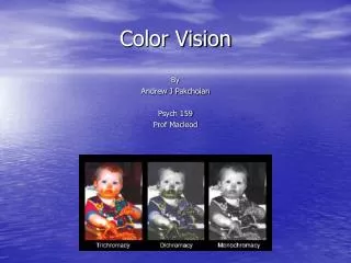
Color Vision. By Andrew J Pakchoian Psych 159 Prof Macleod. Road Map. Pigment anatomy Photoreceptor anatomy Neurobiology of color vision Physiology of color blindness. Key Terms. MC layer magnocellular PC layer parvocellular LGN lateral geniculate nucleus.

E N D
Color Vision By Andrew J Pakchoian Psych 159 Prof Macleod
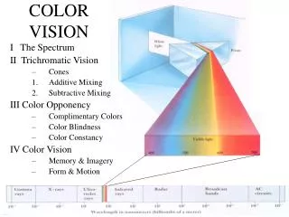
Road Map • Pigment anatomy • Photoreceptor anatomy • Neurobiology of color vision • Physiology of color blindness
Key Terms • MC layer magnocellular • PC layer parvocellular • LGN lateral geniculate nucleus
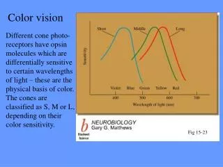
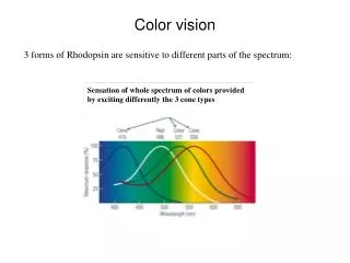
3 types of cones: short (S), middle (M), and long (L) wavelength sensitive. (S): 430 nm = blue (M): 530 nm = green (L): 560 nm = red Pigment Anatomy
Pigment Anatomy • Origin of pigments • Red/green from opsin gene on X-chromosome or sex chromosome. • Show very similar amino acid seqs. (96%) • Blue on chromosome 7 and rhodopsin on chromosome 3 are very different.
Photoreceptor Anatomy • Cones more concentrated near fovea • Adapts to a wide range of illumination colors and levels. • Rods spread throughout the retina • Provide quick response to changes in illumination.
Measuring Wavelengths: Short wavelengths causes the green receptor to fire. As the wavelength gets longer and closer to 580 nm the red begins to fire, surpassing the green. Get mix of wavelengths. Therefore, color vision is the consequence of unequal stimulation of the 3 types of cones. In a specific ratio. Photoreceptor Anatomy
Photoreceptor Anatomy • Example: if you stimulate all 3 types of cones about equally the result is white or no color.
Neurobiology of color vision • Once again 3 types of cones: S, M, and L. • Only 2 types of horizontal cells: • H1 which connects L and M cells. • H2 which connects S with some L and M cells. • The relation between high specificity of cone connectivity and chromatic processing is unclear.
Neurobiology of color vision • Blue/Yellow Pathway: • 2 Systems: • Differentiate signal from S and the summed signal from L and M. = +S-(L+M) • Receives input from S-cone bipolars (ON) and L and M cone bipolars (OFF) • Second system = -S+(L+M) • Input signals are unclear. • Conclusion: The ganglion cells : Small bistratified cell and small bodied inner cell form blue/yellow pathway.
Neurobiology of color vision • Red/Green Pathway: • Differentiate between signals from the L and M cones. • Path: Single L and M cones receive input connect to single midget bipolar cells contact single midget ganglion cells project to the PC layer of LGN.
Neurobiology of color vision • Red/Green Pathway Cont’d: • L and M cones have two forms: ON and OFF-centre • ON-center midget cells for L and M have identical arrays and same with the OFF-center • There is no overlap between dendritic trees of central midget ganglion cells which leads to the exact 1:1 ratio of cones to ganglion cells. • More than 10 to 15 degrees in the periphery we do find some overlap of dendritic trees in the midget bipolar cells, but not ganglion.
Neurobiology of color vision • Interesting facts: • In the periphery of the retina chromatic sensitivity drastically decreases, but responsiveness of PC cells is unchanged Connectivity to midget bipolar cells is not random.
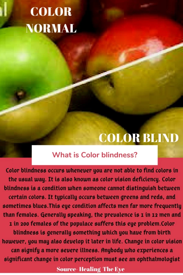
Physiology of color blindness • Male dominant trait but females carry it. • Females have 2 X chromosomes so trait is normally not expressed. • Males have 1 X and 1 Y chromosome which means recessive traits will show in phenotype. • As stated before, red and green pigments originate on the X chromosome. • 1 in 20 males suffers from some form of color blindness. • Most common is red-green color blindness which is caused by problem with M or L cones. • However, they can still see red and green, but have trouble with light or desaturated colors.
Physiology of color blindness • A few types: • Anomalous Trichromacy: have 3 photopigments, but only from 2 groups. • Most common is deuteranomalous = 2 L photopigments. • Dichromacy: missing 1 group of photopigments.
Quiz True or false: The dendritic trees of midget ganglion cells overlap.
Answer False
References • Colorblindness: http://srv2.lycoming.edu/~newman/courses/bio22298/disorderpapers/Colorblindness/preliminary.htm • Hubel, David H. Eye, Brain, and Vision. Harvard Medical School. 15 Jan.2005 • Lee, Barry B. "Paths to Colour in the Retina." Rev. of Current understandingof neurobiology of color vision. Clinical and Experimental Optometry20 June 2004: 239-248. • Levine, Michael W. Fundamentals of Sensation and Perception. 3rd ed. NewYork: Oxford, 2000. 94-110. • Neitz, Jay , and Maureen Neitz. Neitz Color Vision Lab. 21 Feb. 2002.Dept. of Opthamology, Medical College of Wisconsin. 14 Jan. 2005. • Scott, Ed. Spectral Selectivity. 1997. Luminal Path Corporation and contributors. 15Jan. 2005.