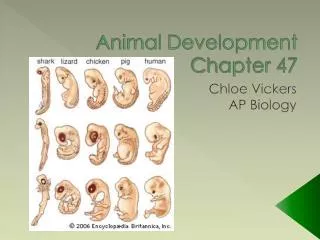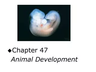
CHAPTER 43 Animal Development
CHAPTER 43 Animal Development. Chapter 43: Animal Development. Fertilization: Interactions of Sperm and Egg Cleavage: Repackaging the Cytoplasm Gastrulation: Producing the Body Plan. Chapter 43: Animal Development. Neurulation: Initiating the Nervous System Extraembryonic Membranes

CHAPTER 43 Animal Development
E N D
Presentation Transcript
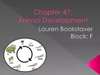
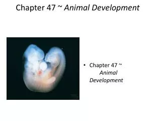
Chapter 43: Animal Development Fertilization: Interactions of Sperm and Egg Cleavage: Repackaging the Cytoplasm Gastrulation: Producing the Body Plan
Chapter 43: Animal Development Neurulation: Initiating the Nervous System Extraembryonic Membranes Human Pregnancy and Birth
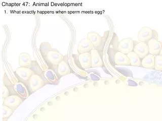
Fertilization: Interactions of Sperm and Egg • Fertilization involves sperm activation • the acrosomal reaction • digestion of a path thru outer egg covering • species-specific binding of sperm to outer egg covering • fusion of sperm and egg cell membranes. Review Figure 43.1 4
figure 43-01.jpg Figure 43.1 Figure 43.1
Fertilization: Interactions of Sperm and Egg • Entry of sperm into egg triggers fast and slow blocks to polyspermy, and in mammals, signals the egg to complete meiosis and begin development. Review Figures 43.2, 43.3, 43.4 6
figure 43-02.jpg Figure 43.2 Figure 43.2
figure 43-03.jpg Figure 43.3 Figure 43.3
figure 43-04.jpg Figure 43.4 Figure 43.4
Fertilization: Interactions of Sperm and Egg • Sperm and egg contribute differentially to the zygote. • The sperm contributes a haploid nucleus and, in some species, a centriole. • The egg contributes a haploid nucleus, nutrients, ribosomes, mitochondria, and informational molecules that will control early stages of development. 10
Fertilization: Interactions of Sperm and Egg • The cytoplasmic contents of the egg are not distributed homogeneously, and are rearranged after fertilization to set up the major axes of the future embryo. Review Figures 43.5, 43.6 11
figure 43-05.jpg Figure 43.5 Figure 43.5
figure 43-06a.jpg Figure 43.6 – Part 1 Figure 43.6 – Part 1
figure 43-06b.jpg Figure 43.6 – Part 2 Figure 43.6 – Part 2
Cleavage: Repackaging the Cytoplasm • In most animals, cleavage is a period of rapid cell division without cell growth or gene expression. • During cleavage, the cytoplasm of the zygote is repackaged into smaller and smaller cells. 15
Cleavage: Repackaging the Cytoplasm • Cleavage pattern is influenced by amount of yolk impeding cleavage furrow formation and orientation of mitotic spindles. • The result of cleavage is a mass of cells called a blastula. Review Figure 43.7 16
figure 43-07a.jpg Figure 43.7 – Part 1 Figure 43.7 – Part 1
figure 43-07b.jpg Figure 43.7 – Part 2 Figure 43.7 – Part 2
Cleavage: Repackaging the Cytoplasm • Cleavage in mammals is unique in that cell divisions are much slower and genes are expressed early in the process. • Cleavage results in an inner cell mass that becomes the embryo and an outer cell mass that becomes the trophoblast. • The mammalian embryo at this stage is called a blastocyst. Review Figure 43.8 19
figure 43-08.jpg Figure 43.8 Figure 43.8
Cleavage: Repackaging the Cytoplasm • Fate maps, which identify what tissues and organs will form from particular blastomeres, can be created for the blastula. Review Figure 43.9 21
figure 43-09.jpg Figure 43.9 Figure 43.9
Cleavage: Repackaging the Cytoplasm • Some species undergo mosaic development: the fate of each cell is determined by the 8-cell stage. • Other species undergo regulative development: cells are not determined so early and can change developmental fates. • In these species, blastomeres separated at early stages can develop into complete embryos, which are then monozygotic, or identical, twins. Review Figure 43.10 23
figure 43-10a.jpg Figure 43.10 – Part 1 Figure 43.10 – Part 1
figure 43-10b.jpg Figure 43.10 – Part 2 Figure 43.10 – Part 2
Gastrulation: Producing the Body Plan • Gastrulation involves massive cell movements that produce three primary germ layers and place cells from various regions of the blastula into new associations with one another. Review Table 43.1 26
table 43-01.jpg Table 43.1 Table 43.1
Gastrulation: Producing the Body Plan • The initial step of sea urchin and amphibian gastrulation is inward movement of certain blastomeres. • The site of inward movement becomes the blastopore. • Cells that move into the blastula become the endoderm and mesoderm; cells remaining on the outside become the ectoderm. • Cytoplasmic factors in the vegetal pole cells are essential to initiate development. Review Figure 43.11, 43.12 28
figure 43-11.jpg Figure 43.11 Figure 43.11
figure 43-12.jpg Figure 43.12 Figure 43.12
Gastrulation: Producing the Body Plan • Gastrulation in frogs is initiated when cells in the gray crescent move into the blastocoel. • This inward migration creates the blastopore. • The dorsal lip of the blastopore is a critical site for the determination of tissues. • It has been called the primary embryonic organizer. Review Figures 43.13, 43.14, 43.15 31
figure 43-13a.jpg Figure 43.13 – Part 1 Figure 43.13 – Part 1
figure 43-13b.jpg Figure 43.13 – Part 2 Figure 43.13 – Part 2
figure 43-14.jpg Figure 43.14 Figure 43.14
figure 43-15a.jpg Figure 43.15 – Part 1 Figure 43.15 – Part 1
figure 43-15b.jpg Figure 43.15 – Part 2 Figure 43.15 – Part 2
Gastrulation: Producing the Body Plan • The anterior–posterior axis of the frog blastula appears to be determined by the distribution of the protein -catenin • This activates a signaling cascade that induces the primary embryonic organizer. 37
Gastrulation: Producing the Body Plan • Gastrulation in reptiles and birds differs from that in sea urchins and frogs because the large egg yolk causes the blastula to form a flattened disc of cells. Review Figure 43.16 38
figure 43-16a.jpg Figure 43.16 – Part 1 Figure 43.16 – Part 1
figure 43-16b.jpg Figure 43.16 – Part 2 Figure 43.16 – Part 2
Gastrulation: Producing the Body Plan • Mammals have a pattern of gastrulation similar to that of birds, even though they have no yolk. Review Figure 43.17 41
figure 43-17.jpg Figure 43.17 Figure 43.17
Neurulation: Initiating the Nervous System • Neurulation follows gastrulation. • Cells that migrate over the dorsal lip of the blastopore are determined to be chordomesoderm, which forms the notochord. • The notochord induces the overlying ectoderm to thicken, form parallel ridges, and fold in on itself to form a neural tube below the epidermal ectoderm. • The nervous system develops from the neural tube. Review Figure 43.18 43
figure 43-18a.jpg Figure 43.18 – Part 1 Figure 43.18 – Part 1
figure 43-18b.jpg Figure 43.18 – Part 2 Figure 43.18 – Part 2
figure 43-18c.jpg Figure 43.18 – Part 3 Figure 43.18 – Part 3
Neurulation: Initiating the Nervous System • The notochord and neural crest cells participate in the segmental organization of tissues called somites along the body axis. • Rudimentary organs and organ systems form during this stage. Review Figure 43.19 47
figure 43-19.jpg Figure 43.19 Figure 43.19
Neurulation: Initiating the Nervous System • Four families of Hox genes determine anterior–posterior pattern differentiation along the body axis in mammals. • Other genes such as sonic hedgehog, contribute to dorsal–ventral differentiation. Review Figure 43.20 49
figure 43-20a.jpg Figure 43.20 – Part 1 Figure 43.20 – Part 1