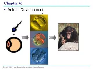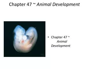
Chapter 47 Animal Development
Chapter 47 Animal Development. Nicole Gallup. Embryonic Development. Genomes of zygote and differences btwn early embryonic cells determine development Cytoplasmic Determinants – Uneven distribution of maternal substances in the unfertilized egg

Chapter 47 Animal Development
E N D
Presentation Transcript
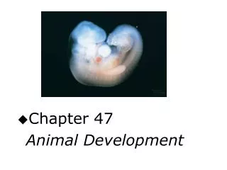
Chapter 47 Animal Development Nicole Gallup
Embryonic Development Genomes of zygoteand differences btwn early embryonic cells determine development Cytoplasmic Determinants – Uneven distribution of maternal substances in the unfertilized egg Differences between cells because of their location in the embryo Cell Differentiation– specialization of cells form and function, caused by gene expression Morphogenesis – process by which an embryo takes shape and cells are in the appropriate locations
Embryonic Stages • Fertilization – When Gametes (sperm and egg) unite • Cleavage – Rapid Cell divisions after Fertilization. S phase (DNA synthesis) and M phase (mitosis). Skips protein synthesis • Gastrulation – Morphogenetic phase Drastic rearrangement of the cells of the blastula. Forms a three-layered embryo with a primitive gut. • Organogenesis – When regions of the three-layered embryo develop into fundamental organs
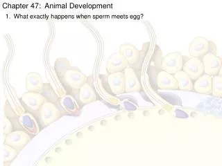
Fertilization Vocab • Acrosomal Reaction - discharge of a sperm’s acrosome when it is near the egg • Acrosome – Vesicle at the tip of sperm, helps sperm penetrate the egg • Fast Block to Polyspermy – Depolarization of egg membrane after sperm binds to vitelline layer. Prevents more sperm from entering • Fertilization Envelope - the changed vitelline layer – prevents other sperm from entering the egg • Slow Block to Polyspermy – Formation of fertilization envelope and other changes, opposite of Fast block, lasts longer
Fertilization • Fertilize externally – eggs and sperm are released at the same time. • Sperm touches egg’s jelly coat – triggering release of acrosome – hole is formed in jelly • Acrosomal process forms – protrudes from sperm, penetrates jelly coat, binds to receptors on egg cell – aka acrosomal reaction • Hole made in vitelline layer – allows contact and fusion of gamete plasma membranes – membranes depolarize forming Fast block • Sperm nucleus enters cytoplasm of egg – then slow block forms
Cleavage Vocab • Blastomer – smaller cells that the embryo divides into • Morula – cluster of cells after the first 5-7 divisions • Blastocoel – a fluid filled cavity • Blastula – hollow ball of cells • Yolk – stored nutrients – distributed differently in all embryos • Vegetal Pole – The pole that the yolk is most concentrated • Animal Pole – Opposite pole, very little yolk
Cleavage • After fusion of gametes cytoplasm rearranges forming 1 body axis. Other axes form later • First 2 divisions are meridional (Vertical) = 4 blastomers of equal size • Third division is equatorial (Horizontal) = 8 blastomers of unequal size – Animal hemisphere = small cells, Vegetal hemisphere = lager cells • Blastula is located in the Animal Hemisphere
Gastrulation Vocab • Gastrula – 3 layered Embryo • Germ Layers – The 3 layers produced. • Ectoderm – Outer layer • Endoderm – Inner Layer • Mesoderm – Partly fills space between Ecto and Endo • Invagination – When cells fold inward • Archenteron – Primitive Gut • Blastopore – Opening in the archenteron, develops into the anus.
Gastrulation • Complicated mechanics – Large amount of yolk & blastula is more than 1 cell thick • Begins on back side of Blastula – cells begin to invaginate in the line along the region • Dorsal Lip – The Dorsal side of the blastopore • Lip extends and invagination continues until the two ends on the blastopore meet on the ventral side • Involution – When future endoderm and mesoderm cells on the surface roll over edge of the lip into the interior of the embryo
Gastrulation • Inside – cells move away from blastopore and become germ layers and blastocoel collapses • Yolk Plug – Large food-laden endodermal cells surrounded by blastopore • End of Gastrulation, circular lip of blastopore encircles plug, cells on surface becomes the ectoderm • Anus forms from the blastopore and mouth develops at the opposite end.
Organogenesis Vocab • Notochord – Formed from dorsal mesoderm • Neural Tube – when neural plate curves inward – rolling into itself • Neural Crest – band of cells along border of Neural tube • Somites – Paired blocks of mesoderm lateral to notochord
Organogenesis • First organs to take shape – neural tube and notochord • Signals from notochord to ectoderm cause ectoderm to become neural plate • Cells from neural crest migrate to all parts of the body – form peripheral nerves, teeth, skull bones • Some somites become wandering cells – go to new locations. • Organogenesis continues – cell differentiation continues to refine organs
Neural Plate formation Neural Tube Formation Somites
Morphogenesis • Major aspect of development in animals – involves movement of cells. • Changes in shape involve reorganization of the cytoskeleton. Cytoskeleton drives cell migration. • Cells that move 1st drag others behind them – directs movement of a sheet if cells • Convergent Extension – morphogenetic movement – cells of tissue layer rearrange, sheets become narrow (converge) and become longer (extend)
Extracellular Matrix • Extracellular Matrix (ECM) – Mixture of secreted glycoproteins outside plasma membrane of cells – trigger/guide movement • Some ECMs promote migration, providing specific molecular anchorage for moving cells • Others keep cells on correct paths – inhibiting migration – use nonmigratory cells • Cell Adhesion Molecules (CAMs) – glycoproteins – help cell migration and stable tissue structure • Cadherins – important cell-to-cell adhesion molecule.
Developmental Fate of Cells • Development requires a combo of morphogenetic changes and the timely differentiation of cells in specific location • 2 general principles • Early cleavage divisions – Embryonic cells must become different from each other • Once initial cells asymmetries are set up, subsequent interactions among the embryonic cells influence their fate – usually causing changes in gene expression
A Cell’s Fate • Fate Maps – diagram of embryonic development – reveals future development of individual cells/tissues • A cell’s fate can be changed by moving the cell to a new location • 2 Important conclusions • Specific tissues of the older embryo can be attributed to certain early “founder cells” • As development proceeds a cell’s developmental Potential becomes restricted
Establishing Cellular Asymmetries • Establishing basic body plan is 1st step in morphogenesis – a prerequisite for the development of tissues/organs • Totipotent – describes a cell that can become any part of an organism • Zygote’s pattern of cleavage affects the fate of cells • Progressive restriction of potency is a feature of development in all animals • The tissue-specific fates of cells in late gastrula are fixed
Inductive Signals • Cell division creates cells that differ from each other the cells then influence each other’s fate (induction) • Pattern Formation – development of an animal’s spatial organization, arrangement of organs/tissues – influenced by inductive signals • Positional Information – Molecular cues – control pattern formation
Limbs • Limbs begin as bumps of tissue called Limb buds • Buds – consist of a core of mesoderm tissue covered by a layer of ectoderm – 2 organizer locations affect limb’s development • Apical Ectodermal Ridge (AER) – 1 organizer – thickened area of ectoderm at the tip of the bud • Zone of Polarizing Activity (ZPA) – other organizer – block of mesodermal tissue located underneath ectoderm – posterior side of the bud is attached to body
Citations • http://www.vcharkarn.com/uploads/0/80.jpg • http://3.bp.blogspot.com/_NDw_XebDkYI/S7ApTP1gibI/AAAAAAAAAO4/aYitNrMkyWo/s1600/cleavage.jpg • http://bio1152.nicerweb.com/doc/class/bio1152/Locked/media/ch47/47_12FrogGastrulation.jpg • http://bio1151.nicerweb.com/Locked/media/ch47/47_14FrogOrganogenesis_CL.jpg