Download

1 / 61
620 likes | 931 Views
Chapter 16: Basal Ganglia. Outline. Anatomy Circuitry Clinical concepts Clinical cases. Overview of function. BG Influence descending motor systems by participating in complex networks Do not directly project to periphery Lesions lead to two broad phenotypes Hypokinetic Hyperkinetic.

E N D
Outline • Anatomy • Circuitry • Clinical concepts • Clinical cases
Overview of function • BG Influence descending motor systems by participating in complex networks • Do not directly project to periphery • Lesions lead to two broad phenotypes • Hypokinetic • Hyperkinetic
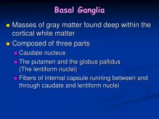
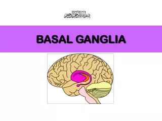
Anatomy • Grey matter nuclei deep within white matter
Anatomy • Relationships • Head of caudate + lentiform nucleus separated by Anterior limb of internal capsule • Lentiform nucleus and thalamus separated by Posterior limb internal capsule • So: • Caudate and thalamus always medial to IC • Putamen and GP always lateral to IC
Brief review vascular supply • Main supply via lenticulostriate branches of MCA • Recurrent artery of Heubner (ACA) • Head of caudate and anterior lentiform nucleus • Anterior choroidal artery (ICA) • Medial GP
Inputs • Main input to BG is to striatum • Cortex +, Glutamate • SNpc + and -, DA • Thalamus +, Glutamate • Intralaminar nuclei in internal medullary lamina • Raphe nuclei modulation, serotonin
Outputs • SNpr and GPi are main outputs, inhibitory • Thalamus • VL, VA, intralaminar, mediodorsal • Pontomedullary reticular formation • Affects reticulospinal tract • Superior colliculus • Affects tectospinal pathways
Intrinsic connections • Excitatory/inhibitory connections within BG • Two main pathways: • Direct pathway • Indirect pathway • Both pathways exert effect via thalamus • Both receive input from SNpc (DAergic nigrostriatal pathway)
Parallel BG pathways • Motor channel • Regulation of movement • Oculomotor channel • Regulation of eye movements • Prefrontal channel • Cognitive processes involving frontal lobes • Limbic channel • Limbic regulation of emotions and motivational drives • Role in neurobehavioral and psychiatric disorders
Understanding movement disorders based on Pathways • Parkinson’s disease • Degeneration of DA-ergic neurons in SNpc • Hemiballismus • Contralateral STN lesion • Huntington’s disease • Degeneration of striatum, initially indirect pthway
Key Clinical Concept 1 –Movement Disorders • “Movement disorders” usually refers to abnormal movement 2ary to BG lesion • Exert effect via projections to cortex • Gradient of hypo- to hyperkinetic
Bradykinesia, Hypokinesia, Akinesia • BG lesions that increase inhibition of thalamus • Loss of DA-ergic input from SNpc • Typified by PD • Loss of inhibition of SNr/GPi via direct pthwy • Loss of inhibition of STN • Other lesions • Diffuse lesions of frontal cortex, subcortical white matter, thalami, reticular formation • Depression, schizophrenia
Rigidity • Increased resistance to passive limb mvment • Spasticity • Velocity-dependent rigidity in UMN lesions • Clasp-knife spasticity (corticospinal tract lesion) • Increase then decreased in resistive tone as stretch • Lead-pipe rigidity (BG lesion) • Continuous resistive tone throughout stretch • Cogwheel rigidity (PD and related disorders) • Rigidity with superimposed tremor • Paratonia or gegenhalten (frontal lobe dysfctn) • Active resistance to limb movement
Dystonia • Abnormal, distorted position of limbs, trunk, face • May be focal, unilateral or generalised • Usually, no focal lesion in BG is found • Examples • Torticollis, blepharospasm, spasmodic dysphonia, writer’s cramp • Etiology • Antipsychotic or anti-emetic medications • Acute or after long-term use (tardivedyskinesia) • BG lesions 2ary infarct, tumor, abscess • Wilson’s, Huntington’s, Parkinson’s disease
Athetosis • Writhing, twisting movements of limbs, face, trunk • May merge with chorea choreoathetosis • Etiology • Perinatal hypoxia involving BG, Kernicterus • Wilson’s disease • Ataxia telangiectasia • Huntington’s disease • Antipsychotic and anti-emetic medications • Treatment of PD with levodopa
Chorea • Nearly continuous, involuntary movements that have fluid or jerky, varying quality • Involves limbs, trunk, neck, face, resp muscles • Range from mild low-amplitude to severe, large-amplitude movements • Worsen with distraction and ambulation • Etiology • Huntington’s disease • Benign familial chorea • Sydenham’s chorea • Lupus • Antipsychotics, anti-emetics, levodopa in PD, phenytoin
Ballismus • Movements of proximal limb muscles with large amplitude, more rotatory or flinging than chorea • Hemiballismus • Unilateral movements 2ary to contralateral BG lesion • Classically, lacunar infarct in STN • Other causes: hemorrhage, tumor, infection
Tics • Sudden, brief action • Preceded by urge to perform action • Followed by sense of relief • Motor tics – usu. involve face, neck • Vocal tics – variable, can be elaborate • Etiology • Tourette’s syndrome • Idiopathic tic disorders • 2ary to encephalitis, infarcts, hemorrhage, tumors
Myoclonus • Sudden, rapid muscular jerk that is focal, unilateral or bilateral • Localisation: cortex, cerebellum, BG, brainstem, spinal cord • Etiology • Anoxic brain injury, encephalitis, toxic and metabolic encephalopathies • Epileptic cortical activity (e.g. JME) • Paraneoplasticdisroders (SCLC, ovarian, breast) • CJD, CBD, late in Alzheimer’s
Tremor • Rhythmic or semi-rhythmic oscillating mvmnts • Both agonist and antagonist muscles involved • Classification • Resting • Parkinsonian (3-5Hz), cerebellar (rubral), palatal • Postural • When pt’s limbs actively held in a position • Essential tremor (5-8Hz), toxic/metabolic, physiologic, NM disorder, PD, cerebellar • Intention (ataxic) • Appears when trying to move limb towards target • Cerebellarappendicular ataxia, postural tremor
Key Clinical Concept 2 – Idiopathic Parkinson’s Disease • Sporadic, unknown etiology, onset 40-70 y.o. • Rarely, familial • Loss of DA-ergic neurons in SNpc • Lewy bodies in remaining DA-ergic neurons • Loss of pigmented neurons elsewhere in CNS • Diagnosis based on clinical features
Idiopathic PD – Clinical Features • Resting tremor • Bradykinesia/hypokinesia • Masked facies • Hypophonia • Slow saccades • Micrographia • Cogwheel rigidity • Postural instability • Retropulsion • Parkinsonian gait • Slow, shuffling, en bloc turn, decreased arm swing • Dementia • 15-40%, late • Bradyphrenia • Starts unilaterally • Insidious progression • Responds to levodopa
L-dopa in Idiopathic PD • Most effective drug for treatment • If no response to treatment consider alternative diagnosis • As disease progresses, on-off phenomena • Abnormal regulation of DA levels • May manage with SR, COMT inhibitors, MAOIs • Other treatment options • DA agonists (e.g. pramipexole) • Anticholinergics (e.g. benztropine) • MAOI (e.g. selegiline)
Parkinsonism Plus Syndromes • Neurodegenerative conditions w/ atypical parkinsonism • Symmetrical Sx, no resting tremor, early postural instability, little response to DA-ergic agents • Examples • Multisystem atrophy • Progressive supranuclear palsy • Lewy body dementia • Cortical basal ganglionic degeneration • Huntington’s, Wilson’s
Huntington’s Disease • Autosomal dominant inheritance pattern • Gene on chromosome 4, CAG trinuc repeats • 4-5/100,000, usu age of onset 30-50 y.o. • Progressive atrophy of striatum • Caudate > putamen > Nacc • All 4 functions of BG affected • Cortical atrophy later in course • Diagnosis • Clinical features, family Hx, genetic testing
Clinical Features • Motor control • Clumsiness, subtle chorea, athetosis, tics, dystonic posturing • Eye movement control • Slow saccades, impaired smooth pursuit, difficulty initiating saccades • Cognition • Decreased attn, decreased memory, impaired executive function • Emotion regulation • Depression, anxiety, OCD, manic-like behavior
Case 1 • 65M, HIV positive • HPI • Involuntary flinging mvmnts of Rt arm/leg • Worsened over 1 mo • Difficult to walk and use hand • O/E • Continuous wild, uncontrollable flapping and circular movements of Rt arm • Jerky mvments of Rt leg • Unsteady gait, falling to right
Case 1 • What would you call this movement disorder? • On the basis of the symptoms and signs, where is the lesion? • What is the most likely diagnosis and what are some possibilities?
Case 1 • Decreased inhibition of contralateral thalamus • Subthalamic lesion • Damage to indirect pathway
Case 2 • 35M, recent-onset jerky mvmnts, marital problems • HPI • Occasional irregular jerking of head, trunk, limbs over past months • Occasional stumbling with fall down stairs • Bitter arguments with wife • Pt denied involuntary mvmnts, gait change, mood disturbance • Family History
Case 2 • Mental status: • Normal orientation, speech, memory. • Blunted affect • CN: • Slow saccades • Motor: • Rare, brief, irregular mvments face, neck, trunk • Slightly decreased tone • Reflexes • Normal and symm • Coordination • Normal • Gait • Unsteady tandem
Case 2 • On the basis of SSx, which of the four channels through BG are involved? • Which part of BG could lead to this disorder? • What is most likely diagnosis? • What genetic abnormality causes this disorder? • What parts of the brain are predominantly affected?
Case 3 • 53M, Rt-handed, 2nd opinion for bradykinesia, tremor, rigidity, unsteady gait • HPI • 10 yrs ago, slowness and difficulty using Rt arm • 8 yrs ago shaking of Rt arm and leg • Dx PD Sinemet started, beneficial • Progressively worse Sx • Tremor of whole body, slower, stiffer, difficulty initiating movements. • Family Hx negative, no antipsychotic meds
Case 3 • Mental status • Micrographia • CN • Mask-like facies, hypophonia • Motor • 4Hz tremor U+L/E, Rt > Lt • Cogwheel rigidity • FFM, RAM slow • Reflexes • No extinction of glabellar (+ve Myerson) • Coordinationn • Slow, no ataxia • Gait • Can’t raise from chair w/o assistance • Slow, stiff gait, stooped posture, short steps, decreased arm swing • En bloc turning
Case 3 • Idiopathic or atypical parkinsonism? • Degeneration of neurons in which structure 1arily responsible for idiopathic PD? • What is main neurotransmitter? • How does loss of neurons lead to hypokinetic disorder? • If pt has on-off phenomena, what procedures can be recommended?