Download
1 / 1
400 likes | 1.38k Views
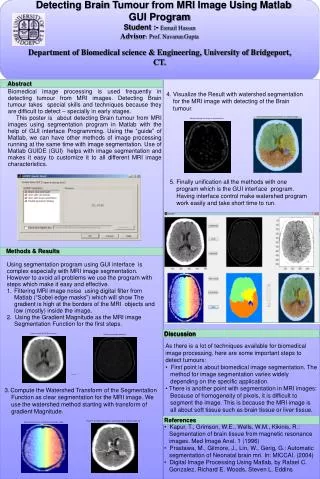
Detecting Brain Tumour from MRI Image Using Matlab GUI Program Student :- Esmail Hassan Advisor : Prof. Navarun Gupta Department of Biomedical science & Engineering, University of Bridgeport, CT. Abstract.

E N D
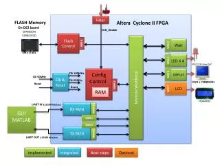
Detecting Brain Tumour from MRI Image Using Matlab GUI Program Student :- Esmail Hassan Advisor: Prof. Navarun Gupta Department of Biomedical science & Engineering, University of Bridgeport, CT. Abstract Biomedical image processing is used frequently in detecting tumour from MRI images. Detecting Brain tumour takes special skills and techniques because they are difficult to detect – specially in early stages. This poster is about detecting Brain tumour from MRI images using segmentation program in Matlab with the help of GUI interface Programming. Using the “guide” of Matlab, we can have other methods of image processing running at the same time with image segmentation. Use of Matlab GUIDE (GUI) helps with image segmentation and makes it easy to customize it to all different MRI image characteristics. 4. Visualize the Result with watershed segmentation for the MRI image with detecting of the Brain tumour. 5. Finally unification all the methods with one program which is the GUI interface program. Having interface control make watershed program work easily and take short time to run. Methods & Results Using segmentation program using GUI interface is complex especially with MRI image segmentation. However to avoid all problems we use the program with steps which make it easy and effective. Filtering MRI image noise using digital filter from Matlab (“Sobel edge masks”) which will show The gradient is high at the borders of the MRI objects and low (mostly) inside the image. 2. Using the Gradient Magnitude as the MRI image Segmentation Function for the first steps. Discussion • As there is a lot of techniques available for biomedical image processing, here are some important steps to detect tumours: • First point is about biomedical image segmentation. The • method for image segmentation varies widely • depending on the specific application. • There is another point with segmentation in MRI images: • Because of homogeneity of pixels, it is difficult to • segment the image. This is because the MRI image is • all about soft tissue such as brain tissue or liver tissue. 3.Compute the Watershed Transform of the Segmentation Function as clear segmentation for the MRI image. We use the watershed method starting with transform of gradient Magnitude. References • Kapur, T., Grimson, W.E., Wells, W.M., Kikinis, R.: • Segmentation of brain tissue from magnetic resonance • images. Med Image Anal. 1 (1996) • Prastawa, M., Gilmore, J., Lin, W., Gerig, G.: Automatic • segmentation of Neonatal brain mri. In: MICCAI. (2004) • Digital Image Processing Using Matlab, by Rafael C. • Gonzalez, Richard E.Woods, Steven L. Eddins