Download
1 / 3
50 likes | 116 Views
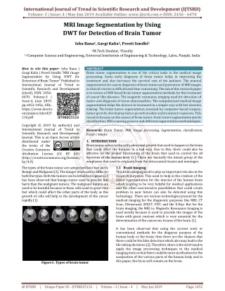
MR image segmentation assumes a significant job and a significant job in the restorative field because of its assortment of utilizations particularly in Brain tumor investigation. Cerebrum tumor is an unusual and uncontrolled development of cells. It occupies room inside the skull. It can pack, move and damage solid cerebrum tissue and nerves. Additionally as a rule it deter with ordinary mind work. Tumors can be kindhearted non dangerous or threatening malignant , can occur in various pieces of the cerebrum. Cerebrum tumor arrangement and ID from Magnetic Resonance MR information is a fundamental. However, it requires some serious energy and manual errand finished by restorative pros. Mechanizing this undertaking is a difficult due to the high assortment in the vibe of tumor tissues among various patients and by and large similitude with the ordinary tissues. Right now, tumor picture has been portioned utilizing proposed Fuzzy grouping calculation FCM . The presentation of FCM division strategy is contrasted and those of watershed and SVM calculations. Pavithra. R | E. Sivaraman "MRI Brain Image Segmentation using Fuzzy Clustering Algorithms" Published in International Journal of Trend in Scientific Research and Development (ijtsrd), ISSN: 2456-6470, Volume-4 | Issue-3 , April 2020, URL: https://www.ijtsrd.com/papers/ijtsrd30316.pdf Paper Url :https://www.ijtsrd.com/engineering/electrical-engineering/30316/mri-brain-image-segmentation-using-fuzzy-clustering-algorithms/pavithra-r<br>

E N D
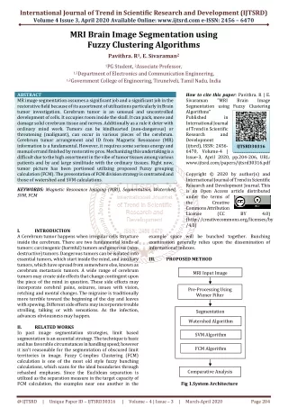
International Journal of Trend in Scientific Research and Development (IJTSRD) Volume 4 Issue 3, April 2020 Available Online: www.ijtsrd.com e-ISSN: 2456 – 6470 MRI Brain Image Segmentation using Fuzzy Clustering Algorithms Pavithra. R1, E. Sivaraman2 1PG Student, 2Associate Professor, 1,2Department of Electronics and Communication Engineering, 1,2Government College of Engineering, Tirunelveli, Tamil Nadu, India ABSTRACT MR image segmentation assumes a significant job and a significant job in the restorative field because of its assortment of utilizations particularly in Brain tumor investigation. Cerebrum tumor is an unusual and uncontrolled development of cells. It occupies room inside the skull. It can pack, move and damage solid cerebrum tissue and nerves. Additionally as a rule it deter with ordinary mind work. Tumors can be kindhearted (non-dangerous) or threatening (malignant), can occur in various pieces of the cerebrum. Cerebrum tumor arrangement and ID from Magnetic Resonance (MR) information is a fundamental. However, it requires some serious energy and manual errand finished by restorative pros. Mechanizing this undertaking is a difficult due to the high assortment in the vibe of tumor tissues among various patients and by and large similitude with the ordinary tissues. Right now, tumor picture has been portioned utilizing proposed Fuzzy grouping calculation (FCM). The presentation of FCM division strategy is contrasted and those of watershed and SVM calculations. KEYWORDS: Magnetic Resonance Imaging (MRI), Segmentation, Watershed, SVM, FCM How to cite this paper: Pavithra. R | E. Sivaraman "MRI Segmentation using Fuzzy Clustering Algorithms" Published in International Journal of Trend in Scientific Research and Development (ijtsrd), ISSN: 2456- 6470, Volume-4 | Issue-3, April 2020, pp.204-206, URL: www.ijtsrd.com/papers/ijtsrd30316.pdf Copyright © 2020 by author(s) and International Journal of Trend in Scientific Research and Development Journal. This is an Open Access article distributed under the terms of the Creative Commons Attribution License (CC (http://creativecommons.org/licenses/by /4.0) Brain Image IJTSRD30316 BY 4.0) I. A Cerebrum tumor happens when irregular cells structure inside the cerebrum. There are two fundamental kinds of tumors: carcinogenic (harmful) tumors and generous (non- destructive) tumors. Dangerous tumors can be isolated into essential tumors, which start inside the mind, and auxiliary tumors, which have spread from somewhere else, known as cerebrum metastasis tumors. A wide range of cerebrum tumors may create side effects that change contingent upon the piece of the mind in question. These side effects may incorporate cerebral pains, seizures, issues with vision, retching and mental changes. The migraine is traditionally more terrible toward the beginning of the day and leaves with spewing. Different side effects may incorporate trouble strolling, talking or with sensations. As the infection, advances obviousness may happen. II. RELATED WORKS In past image segmentation strategies, limit based segmentation is an essential strategy. The technique is basic and has favorable circumstances in handling speed; however it isn't reasonable for the segmentation of obscured limit territories in image. Fuzzy C-implies Clustering (FCM) calculation is one of the most old style fuzzy bunching calculations, which scans for the ideal boundaries through rehashed emphases. Since the Euclidean separation is utilized as the separation measure in the target capacity of FCM calculation, the examples near one another in the INTRODUCTION example space will be bunched together. Bunching examination generally relies upon the dissemination of informational indexes. III. PROPOSED METHOD Fig 1.System Architecture MRI Input Image Pre-Processing Using Wiener Filter Segmentation Watershed Algorithm SVM Algorithm FCM Algorithm Comparative Analysis @ IJTSRD | Unique Paper ID – IJTSRD30316 | Volume – 4 | Issue – 3 | March-April 2020 Page 204
International Journal of Trend in Scientific Research and Development (IJTSRD) @ www.ijtsrd.com eISSN: 2456-6470 A.Pre-Processing Parametric Description The exhibition parameters are most significant criteria to legitimize the reenactment results. Peak signal to noise ratio (PSNR) and mean square error (MSE) are viewed as parameters. The nature of denoised picture is estimated by PSNR = 10 Where is maximum value of pixel present in an image and MSE is the mean square error between original and denoised image with M*N size. MSE = B.Segmentation MR image segmentation is the most fundamental and significant piece of picture handling which portions a picture into important territories as per a few attributes, for example, dark level, range, surface, shading, etc. The objective of image segmentation is to segment a picture into a lot of disjoint districts with uniform and homogeneous qualities, for example, force, shading, tone or surface and so on. C.Watershed segmentation method The watershed based techniques utilizes the idea of topological translation. Right now speaks to the bowls having gap in its minima from where the water spills. At the point when water arrives at the outskirt of bowl the contiguous bowls are consolidated. To keep up detachment between bowls dams are required and are the fringes of district of division. These dams are built utilizing widening. The watershed strategies think about the inclination of picture as topographic surface. The pixels having more angles are spoken to as limits which are nonstop. D.SVM Techniques SVM or Support Vector Machine is a direct model for grouping and relapse issues. It can take care of straight and non-direct issues and function admirably for some commonsense issues. The possibility of straightforward: The calculation makes a line or a hyperplane which isolates the information into classes. E.Fuzzy C-Means algorithm Fuzzy grouping is a type of bunching where every datum point can have a place with more than one bunch. Bunching or group investigation includes appointing information focuses to groups with the end goal that things in a similar bunch are as comparative as could be expected under the circumstances, while things having a place with various bunches are as unique as could be expected under the circumstances. Bunches are distinguished by means of comparability measures. These closeness measures incorporate separation, network, and force. Diverse likeness measures might be picked dependent on the information or the application. IV. RESULTS AND DISCUSSION The unwanted noise present in the brain MR image has been eliminated by using the wiener filter. Table1. Filtered MR brain images (1) . (2) Table2. PSNR values of sample images S. NO IMAGE 1 Sample Image 1 2 Sample Image 2 3 Sample Image 3 MSE PSNR (dB) 7.74 39.2774 4.44 41.6938 3.86 42.2955 Table3. Segmented MR brain images (Watershed) SVM is @ IJTSRD | Unique Paper ID – IJTSRD30316 | Volume – 4 | Issue – 3 | March-April 2020 Page 205
International Journal of Trend in Scientific Research and Development (IJTSRD) @ www.ijtsrd.com eISSN: 2456-6470 Table4. Segmented MR brain images (SVM) outcome is evident that FCM Algorithm delivers better outcome contrasted and those of Watershed and SVM Algorithms as far as calculation time and DSC values. REFERENCES [1]Roerdink, J. B. and Meijster, A., 2000. The watershed transform: Definitions, algorithms and parallelization strategies. Fundamenta informaticae, 41(1, 2), pp.187- 228. [2]Glasbey, C. A. and Horgan, G. W., 1995. Image analysis for the biological sciences (Vol. 1). Chichester: Wiley. [3]Shafarenko, L., Petrou, M. and Kittler, J., 1997. Automatic watershed segmentation of randomly textured color images. IEEE transactions on Image Processing, 6(11), pp.1530-1544. [4]Gonzalez, RC and Woods, RE, 2002. Digital image processing. [5]Dokládal, P., Urtasun, R., Bloch, I. and Garnero, L., 2001, October. Segmentation of 3D head MR images using morphological reconstruction under constraints and automatic selection of markers. In Proceedings 2001 International Conference on Image Processing (Cat. No. 01CH37205) (Vol. 3, pp. 1075-1078). IEEE. [6]Pratt, W.K., 2013. Introduction to digital image processing. CRC press. [7]Clark, M. C., Hall, L. O., Goldgof, D. B., Velthuizen, R., Murtagh, F.R. and Silbiger, M.S., 1998. Automatic tumor segmentation using knowledge-based techniques. IEEE transactions on medical imaging, 17(2), pp.187-201. [8]LIU, Y. T., Zhang, H. X. and Li, P. H., 2011. Research on SVM-based MRI image segmentation. The Journal of China Universities of Posts and Telecommunications, 18, pp.129-132. [9]Guo, L., Liu, X., Wu, Y., Yan, W. and Shen, X., 2007, August. Research on the segmentation of MRI image based on multi-classification support vector machine. In 2007 29th Annual International Conference of the IEEE Engineering in Medicine and Biology Society (pp. 6019-6022). IEEE. [10]Xiao, J. and Tong, Y., 2014, May. Research of Brain MRI image segmentation algorithm based on FCM and SVM. In The 26th Chinese Control and Decision Conference (2014 CCDC) (pp. 1712-1716). IEEE. [11]Kasiri, K., Kazemi, K., Dehghani, M. J. and Helfroush, M.S., 2010, July. Atlas-based segmentation of brain MR images using least square support vector machines. In 2010 2nd International Conference on Image Processing Theory, Tools and Applications (pp. 306- 310). IEEE. [12]Ruan, S., Lebonvallet, S., Merabet, A. and Constans, J. M., 2007, April. Tumor segmentation from a multispectral MRI images by using support vector machine classification. In 2007 Symposium on Biomedical Imaging: From Nano to Macro (pp. 1236-1239). IEEE. [13]Sudhakar, B. and Veluthai, M., Brain Tumor Segmentation of MRI Image using Gustaffson-Kessel (GK) Fuzzy Clustering Algorithm. [14]Xess, M. and Agnes, S. A., 2014. Analysis of image segmentation methods based on performance evaluation parameters. Int J Comput Eng Res, 4(3), pp.68-75. [15]Zhang, Y. J., 1996. A survey on evaluation methods for image segmentation. Pattern pp.1335-1346. Table 5.Segmented MR brain images (FCM) Table6. Performance Analysis Computation Time(Seconds) Watershed 0.0624 SVM 0.0356 FCM 0.0156 DSC Values 0.3692 0.3483 0.1527 Algorithm 4th IEEE International FCM Algorithm produces better result compared with those of Watershed and SVM Algorithms in terms of computation time and DSC values. V. CONCLUSION MR image has been done utilizing Wiener channel. The division has been finished by using Watershed, SVM and FCM Algorithms. The presentation of FCM has been contrasted and those of Watershed and SVM Algorithms. From the recognition, 29(8), @ IJTSRD | Unique Paper ID – IJTSRD30316 | Volume – 4 | Issue – 3 | March-April 2020 Page 206