Download
1 / 37
370 likes | 497 Views
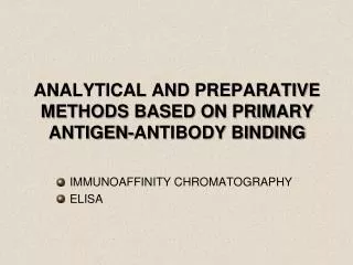
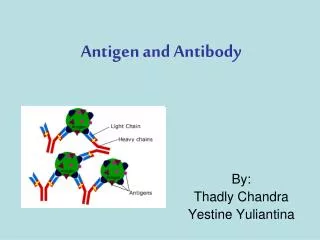
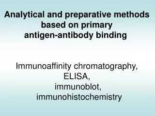
Analytical and preparative methods based on primary antigen-antibody binding Immunoaffinity chromatography, ELISA, immunoblot, immun o histochemistry. Affinity purification of antibodies using an antigen-sorbent column. column. polymer beads. Column content. Covalently bound

E N D
Analytical and preparative methods based on primary antigen-antibody binding Immunoaffinity chromatography, ELISA, immunoblot, immunohistochemistry
Affinity purification of antibodies using an antigen-sorbent column column polymer beads Column content Covalently bound antigen
Addition of antibodies to be purified Binding Washing elution Antigen-specific polyclonal antibodies
The reverse setup is also used column Column content Covalently bound antibodies Purification of antigen on antibody-immunosorbent column polymer beads Loading the antigen mixture Binding Wash Elution
ELISA Enzyme Linked ImmuneSorbent Assay ELISA plate Well
enzyme enzyme linked immune sorbent Antigen/antibody adsorbed to solid surface Antibody conjugated with enzyme
Enzyme activity in ELISA is directly proportional to the amount of antigen present Enzyme activity is measured by the color reaction due to conversion of substrate Similar principle applies to many other antibody-based detection methods
Label Secondary antibodies Label Antigen Basic setups in ELISA, immunohistochemistry, flow cytometry Indirect method Direct method Primary antibodies
Enzyme Enzyme-specific antibody, same isotype as the primary antibody Secondary antibody Primary antibody Antigen Basic setups in ELISA, immunohistochemistry, flow cytometry Enzyme/anti-enzyme system PAP – peroxidase / anti-peroxidase APAAP – alkaline phosphatase / anti- alkaline phosphatase
Avidin-biotin enzyme complexes Avidin-enzymecomplexes Biotin-enzyme complex Avidin Biotinylated antibody Antigen Basic setups in ELISA, immunohistochemistry, flow cytometry Indirect systems combined with biotin-avidin signal amplification (Avidin binds biotin with very high affinity ) ABC Basic
Addition of Secondary Ab conjugated with enzyme Steps of the basic indirect ELISA Detection of antigen or specific antibody Adsorption of antigen (coating) Removal of excess antibody Removal of excess antigen Saturation of uncovered surface area with protein such as BSA, casein etc.. Removal of excess protein Addition of Ag-specific antibodies Addition of chromogenic substrate
Steps of the combined sandwich ELISA For antigens present at low concentration in complex biological samples Removal of excess enzyme Coating with Ag-specific „capture” antibody Removal of unbound material Removal of unbound protein Blocking free plastic surface with inert protein Removal of unbound material Addition of antigen- containing solution Addition of biotinylated antibody specific to a different epitope on target protein Addition of avidin-conjugated enzyme Addition of substrate
Competitive ELISA Highly sensitive method used to detect and quantitate small amounts of antigen in complex biological samples. Antigen in solution and on the solid surface compete for the binding site of labeled specific antibody. - Coat with antigen, blocking - Add experimental sample that contains or lacks antigen -Add labeled antibody, --- Binding - Washes - Add substrate
ELISA Background due to non-specific binding to plastic Labeled antibody bound to plastic directly or indirectly gives the same signal as those that bind specifically • Nonspecific binding can be controlled by: • Saturation of free plastic surfaces • Using detergents
ELISA plates - results The chromogens used for detection are CARCINOGENOUS!
Characterization of antigens by Western blotting
Western Blot • The use of antibodies in molecular biology is widespread • It is probably most often encountered in Western analysis • SDS-PAGE gel resolved into single protein bands (overlap possible) • Presence of a protein is determined by hybridizing the proteins, transferred or applied to a membrane, with the relevant antibody Protein sample Standard Antibody recognizes epitope in specific protein Western blot Membrane SDS-PAGE
Western Blot • Used to detect specific proteins in a sample • Proteins separated by Sodium Dodecyl Sulfate-Polyacrylamide Gel Electrophoresis (SDS-PAGE), transferred to a membrane • Primary (1st) antibody (monoclonal or polyclonal) used to detect protein • Enzyme linked 2nd antibody (e.g. horseradish peroxidase-linked) used to detect 1st antibody
Enhanced chemiluminescence (ECL) • In the presence of H2O2, horseradish peroxidase (HRP) oxidizes diacylhydrazides such as luminol • Directly after oxidation, luminol is in an exited state, and emits a photon to return to the ground state • This photon can be detected with a film or a camera • Light emission can be enhanced by ~1000-fold with phenolic compounds such as 6-hydroxybenzothiazole (enhancer) Immobilized proteins Primary antibody Fab Fc Horseradish peroxidase luminol H2O2 h Epitope on protein surface luminol enhancer Detection H2O Secondary antibody Membrane
Immunohistochemistry Labeled antibodies added to fixed tissue sections detect the distribution of the chosen antigen within the tissue or within the cells of a particular tissue • Immunofluorescence • Fluorescent dye coupled to antibody • FITC – fluorescein isothiocyanate • PE – phycoerythrin • Immunoenzyme method • enzyme-coupled antibody • P – peroxidase • PA – alkaline phosphatase • (Substrates converted into an insoluble compound)
Immunohistochemistry Fixation Sectioning Tissue sample Section before staining Freezing
ImmunohistochemistryABC Method Enzim Avidin X Biotin Secondary antibody Primary antibody Slide Cells Tissue sample
Histochemistry Acute bronchopneumonia (hematoxilin- eozin staining)
Immunohistochemistry (CD68+ macrophages and lymphocytes, granuloma)
Immunohistochemistry using fluorescent detection Anti-nuclear autoantibodies in SLE (FITC)
Immunohistochemistry using fluorescent detection (TRITC) Detection of actin microfilaments
A fixed and permeabilized skin fibroblast.Mitochondria were labeled with mouse IgG (anti–OxPhos Complex V) and visualized using goat anti–mouse IgG conjugated with orange-fluorescent Alexa Fluor 555. F-actinwaslabeled with green-fluorescent Alexa Fluor 488 phalloidin (a mushroom toxin),and the nucleuswas stained with TO-PRO-3 iodide
Peroxisome labeling in fixed and permeabilized pulmonary artery endothelial cell.Peroxisomeswere labeled using an antibody directed at peroxisomal membrane protein 70 and detected with Alexa Fluor 488–labeled goat anti–mouse IgG. Mitochondria were stained with MitoTracker Red prior to fixation; nuclei were stained with blue-fluorescent DAPI.
Acquired Immune Deficiency Syndrome – AIDS Certain infectious microorganisms can supress or subvert the immune system. At the beginning of the last century, when tuberculosis was the leading cause of death and fully half the population was tuberculin-positive, it was well-known that an inter- current measles infection would cause a well-contained tuberculosis infection to run rampant and result in death. The mechanism responsible is now known to be the supression of IL-2 synthesis after binding of measles virus to CD46 on macrophages.
Some of the microorganisms that supress immunity act by infecting lymphocytes. The human immunodeficiency virus (HIV) presents a chilling example of the consequences of infection and destruction of immune cells by a microorganism. The T-cell surface CD4 molecule acts as a receptor for HIV. CD4 is also expressed on the surface of cells of the macrophage lineage and they too can be infected by this virus. The clinical latency is long, usually it means several years. During this period, the level of CD4+ cells and virus particles in the blood changes. When the rate at which CD4+ cells are being destroyed exceeds the capacity of the host to replenish them, their number decreases to a point where cell-mediated immunity falters. The failure of cell-mediated immunity renders to the host susceptible to fatal opportunistic infections.
Case study: The Pinkerton-family: infected blood caused tragedy Benjamin Pinkerton was a US-navy lieutenant who saw service at Japan. He married with a japanese woman during his service, who gave birth two healthy girls in 1987. She bore a boy four years later, who seemed healthy, as well. The boy got the routine DPT-vaccination and an oral polio-virus immunization. These vaccinations had no side-effect and the boy grew normally. At the age of six months he got sick and started to lose weight. He had severe, chronic diarrhea with fever. Besides a chronic oral candidiasis, the boy got two otitis, one after the other . The navy doctors examined the baby several times and prescribed antibiotic but it proved ineffectual. • Results of somatic examination: • body-temperature 38oC; • candidiasis on the lateral sides of tongue • and on the mucosal surface of the oral cavity; • „diaper-pimples”, which is also caused by Candida • infection; • at respiration a subtle, • slurping noise was heard in each pulmonary lobes;
Oral candidiasis esophageal candidiasis
Laboratory: • normal amount of leukocytes (6500/ml); • normal rate of leukocytes (neutrophyl 62%; • lymphocyte 30%; monocyte 5%; eosinophyl 2%; basophyl 1%); • normal serum immunoglobulin levels: • serum IgG: 997 mg/dl (phys.: 800-1000 mg/dl); • serum IgM: 73 mg/dl (phys.: 50-150 mg/dl); • IgA: 187 mg/dl (phys.: 150-300 mg/dl); • normal amount of CD8 + T-cells, but the rate of CD4+ T-cells • is very low, only 85/ml (phys.: 1000-1200/ml); • intradermal Candida-antigen did not evoke late-type • hypersensitvity reaction; • results of ELISA and Western-blot analysis: • HIV-antibodies in the serum;
During HIV-diagnostics samples are always analyzed firstly by ELISA-method. In case of reactivity (serum positive) two more measurements are needed (2nd and 3rd analysis). If the 2nd and 3rd measurements show reactivity as well, the subject’s sample must be verificated: usually a Western blot or another ELISA is performed. After this finding they verified the parents: Both of them were HIV-positive.
While Pinkerton was healthy, his wife was feeling unwell and complained about the swelling of her cervical lymphatic nodes. It turned out, that she was pregnant right before the boy’s birth. At the end of pregnancy the fetus had died and had to be removed by caesarean section. The operation was going well but – because of the loss of blood – she needed blood-transfusion (she got two units of blood). The boy got two severe infections in turn: Pneumocystis carinii- and Pseudomonas aeruginosa. He had serious cough with bloody spit (hemoptysis). A week after this he died. The parents got AZT- (zidovudin) therapy. While his wife died soon in respiratory failure, the lieutenant – in spite of his high serum HIV-antibody level – has not had symptoms yet.