Download
1 / 43
430 likes | 494 Views
Learn about direct and indirect antibody detection, different detection methods for antigen-antibody complexes, and strategies for visible and invisible detection. Discover various techniques like precipitation, agglutination, ELISA, and more. Gain insights into performance, applications, advantages, and limitations of these detection methods.

E N D
Investigation strategies and methods Antigen and antibody detection May 2007
Learning objectives At the end of the presentation, participants should • Understand direct and indirect antibody detection • Understand the different methods for detecting antigens or antibodies
Detection • Detection of antigen-antibody complex • Antigen-antibody complex requires specific conditions • temperature • pH • Complex may be directly visible or invisible
Detection Directly visible – agglutination Invisible • requires specific probes (enzyme-labelled anti-immunoglobulin, isotope-labelled anti-immunoglobulin, etc.) • binds Ag-Ab complex and amplifys signals • signals can be measured by naked eyes or specific equipment e.g. in ELISA, RIA, IFA
Methods for Ag-Ab detection • Precipitation • Agglutination • Hemagglutination and hemagglutination inhibition • Viral neutralization test • Radio-immunoassays • ELISA • Immunoflourescence • Immunoblotting • Immunochromatography
Precipitation Principle • soluble antigen combines with its specific antibody • antigen-antibody complex is too large to stay in solution and precipitates Examples • flocculation test • immuno-diffusion test • counter-immuno-electrophoresis (CIEP)
Flocculation test (precipitation reaction) Principle • precipitate, a concentrate of fine particles, is usually visible (macroscopically or microscopically) because the precipitated product is forced to remain suspended Examples • VDRL slide flocculation test • RPR card test • Kahn’s test for syphilis
Flocculation test (A precipitation reaction) (1) Non Reactive (2) Weakly Reactive (3,4) Reactive RPR card test
Precipitation:Performance, applications • Advantages • sensitive for antigen detection • Limited applications • Time taken - 10 minutes
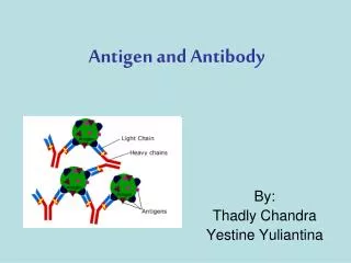
Direct agglutination Ag-Ab complex Positive Negative Principle • combination of an insoluble particulate antigen with its soluble antibody • forms antigen-antibody complex • particles clump/agglutinate • used for antigen detection Examples • bacterial agglutination tests for sero-typing and sero-grouping e.g., Vibrio cholerae, Salmonella spp
Passive (indirect) agglutination Principle • precipitation reaction converted into agglutination - coating antigen onto the surface of carrier particles like red blood cells, latex, gelatin, bentonite • background clears Examples of types • latex agglutination • co-agglutination • passive hemagglutination (treated red blood cells made resistant) Examples of tests - agglutination for leptospirosis Widal test (typhoid fever)
Reverse passive agglutination Principle • antigen binds to soluble antibody coated on carrier particles and results in agglutination • detects antigens Example • detecting cholera toxin
Reverse passive agglutination Positive Negative
Agglutination:Performance, applications Advantages • sensitive for antibody detection Limitations • Prozone phenomenon: • requires the right combination of quantities of antigen and antibody • handled through dilution to improve the match Time taken • 10-30 minutes
Hemagglutination Principle • many human viruses have the ability to bind to the surface structures on red blood cells from different species thereby causing agglutination Example • influenza virus binds to fowl’s red blood cells
Hemagglutination inhibition Principle Antibodies to the virus in the patient serum bind to the virus; blocks binding sites on the viral surfaces • prevents the virus from agglutinating the red cells Example • detecting antibodies to influenza and dengue viruses Positive Negative Hemagglutination inhibition for detection of Dengue antibodies
Hemagglutination:Performance, applications Advantages • highly specific • can be used as gold standard Limitations • technically demanding • time consuming • cannot distinguish IgG from IgM Time taken • 1 day
Neutralization assays Principle • antibodies in serum neutralize antigens on the surface of viruses (neutralizing antibodies) • inhibited viruses cannot infect cell lines Example • plaque neutralization assay for dengue virus, Japanese encephalitis virus • antibodies to bacterial toxins and other extra-cellular products that display measurable activities (e.g., ASLO, diphtheria toxin, clostridium toxin)
Neutralization:Performance, applications • Advantages • Highly specific • Often used as gold standard • Limitations • Technically demanding • Time consuming • Can only be used for viruses that can be grown • Complexity limits the use beyond gold standard • Time taken • 1 week
Radio-immunoassays Response Antibody • Principle • Radioactively labelled-antibody (or antigen) competes with the patient’s unlabelled antibody (or antigen) for binding sites on a known amount of antigen (or antibody) • Reduction in radioactivity of the antigen-patient antibody complex compared with control test is used to quantify the amount of patient antibody / antibody bound • Limited use due to the problems with handling radioisotope • Example • HBsAg • Thyroid function test
Positive Negative Neutralization Assay
Radio-immunoassays:Performance, applications Adantages • highly sensitive • can be used for detection of small quantities • quantification possible Limitations • expensive • requires isotopes Time taken • 1 day
Enzyme-linked immunosorbant assay (ELISA) Labeling technique Principle • use of enzyme-labelled immunoglobulin to detect antigens or antibodies • signals are developed by the action of hydrolyzing enzyme on chromogenic substrate • optical density measured by micro-plate reader Examples • Hepatitis A (Anti-HAV-IgM, anti-HAV IgG)
ELISA Micro-plate reader Positive result Response 96-well micro-plate Antibody
Types of ELISA (Ag Ab tests) Labeling technique Competitive • Antigen or antibody are labelled with enzyme and allowed to compete with unlabeled ones (in patient serum) for binding to the same target • Hydrolysis signal from Ag-Ab complex (enzyme-labelled) is measured • Antigen or antibody in serum is then calculated • No need to remove the excess/unbound Ag or Ab from the reaction plate or tubes)
Types of ELISA used in the detection of antigens and antibodies Labeling technique • Non-competitive • must remove excess/unbound Agor Ab before every step of reactions • Direct ELISA • Indirect ELISA • Sandwich ELISA • Ab Capture ELISA(similar to sandwich ELISA but in 1st step, anti-Ig (M or G) is coated on the plate • Then antibodies in patient serum are allowed to capture in next step
ELISA:Performance, applications • Advantages • Automated, inexpensive • Objective • Small quantities required • Class specific antibodies measurable • Limitations • Expensive initial investment • Variable sensitivity / specificity of variable tests • Cross contamination • Time taken - 1 day
Performance comparison of various ELISAs for antibody detection
Immuno-fluorescence Labeling technique • Principle • Use fluorescein isothiocyanate labeled-immunoglobulin to detect antigens or antibodies according to test systems • Requires a fluorescent microscope • Examples • Herpes virus IgM • Dengue virus • Rabies virus • Scrub and murine typhus Cell infected with Dengue virus V. Cholerae
Immuno-fluorescence:Performance, applications • Advantages • Sensitive and specific • Can be used for discrepant analysis • Limitations • Expensive (Reagents and equipment) • Subjective • Cross reactivity • Non-specific immuno-fluorescence • Time taken • 1 day
Types of immuno-fluorescence Labeling technique • Direct immuno-fluorescence • Used to detect antigen • Indirect and sandwich immuno-fluorescence • Antigen detection • Antibody detection
Western-blot analysis (1) • Principle • Antigens are separated by Poly Acrylomide Gel Electrophoresis (PAGE) and trans-blotted onto nitrocellulose/nylon membranes • Antibodies in serum react with specific antigens • Signals are detected according to the principles of test systems • Antibodies against microbes with numerous cross-reacting antibodies identified more specifically • Examples • T. pallidum, B.burgdorferi, • Herpes simplex virus types 1 and 2 Anti HIV-1
Western-blot analysis (2) • Serum, saliva, urine can be tested • Kits are commercially available • Recombinant immuno-blotting assays (RIBA) uses recombinant proteins Anti HIV-2
Immunoblot:Performance, applications • Advantages • Used for discrepant analysis • Highly specific • Rapid kits available • Limitations • Cost • Concern validated data • Time taken • 1 day
Immuno-chromatography: Principle (1) Lysing agend Labled AB. Control band (bound AB) Test band (bound AB) Bound AB Free labled AB Nitrocellulose strip • Dye-labelled antibody, specific for target antigen, is present on the lower end of nitrocellulose strip or in a plastic well provided with the strip. • Antibody, also specific for the target antigen, is bound to the strip in a thin (test) line • Either antibody specific for the labelled antibody, or antigen, is bound at the control line
Immuno-chromatography: Principle (2) Captured Ag-labelled Ab-complex Captured labelled Ab Labelled AB-AG-complex Captured by bound AB of test band Labelled AB-AG-complex Captured by bound AB of control band • If antigen is present, some labelled antibody will be trapped on the test line • Excess-labelled antibody is trapped on the control line
Immuno-chromatography: Performance, applications • Advantages • Commercially available • Single use, rapid test • Easy to perform • Can detect antigen or antibody • Can be used in the field • Limitations • Cost • Concern validated data • Time taken - 1 hour
Interpretation of antigen detection tests • In general, detection of the antigen denotes a presence of the pathogen • More important in some of parasitic and fungal diseases
Interpretation of two, acute and convalescent IgG tests * * Convalescent serum collected 2-4 weeks after onset
Interpretation of a single IgG test * Collected between onset and convalescence
Elements influencing the sensitivity and specificity of a given test kit • Test format • Precipitation versus IFA, Rapid test versus ELISA • Purity of the antigen used • Crude versus purified antigen versus synthetic peptides • Type of the antibody used • Polyclonal versus monoclonal antibodies • Interfering substances in the sample • Presence of rheumatoid factor in the serum of the patient • Similarity in antigenic composition of pathogens • Cross reactivity
Investigation strategies and methods Developed by:The Department of Epidemic and Pandemic Alert and Response of the World Health Organization with the assistance of:European Program for Field Epidemiology Training Canadian Field Epidemiology Programme Thailand Ministry of Health Institut Pasteur