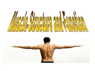
Muscle Structure and Function
Human Physiology Unit. Muscle Structure and Function. Muscle Function. Muscles are for: contraction for locomotion and skeletal movement contraction for propulsion contraction for pressure regulation. Types of Human Muscle.

Muscle Structure and Function
E N D
Presentation Transcript
Human Physiology Unit Muscle Structure and Function
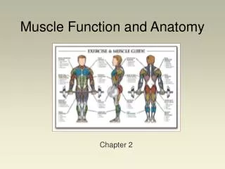
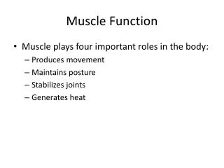
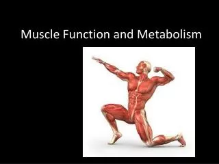
Muscle Function • Muscles are for: • contraction for locomotion and skeletal movement • contraction for propulsion • contraction for pressure regulation
Types of Human Muscle • Based on their structures, contractile properties, and control mechanisms, there are three types of muscle in the human body: • skeletal muscle, • smooth muscle, and • cardiac muscle.
Smooth Muscle • Smooth muscle forms the walls of blood vessels and body organs • Control of is involuntary & by the autonomic nervous system • Contraction is slow & uniform; this muscle is relatively fatigue resistant
Cardiac Muscle • Cardiac muscle makes up your heart • Has characteristics of both skeletal muscle and smooth muscle • Is fatigue resistant & contractions are involuntary
Skeletal Muscle • Skeletal muscle is attached to bone & is responsible for supporting & moving the skeleton • Contraction is under voluntary control by motor neurons.
Skeletal Muscle • Skeletal muscle is a group of muscle fibres bound together by connective tissue • Skeletal muscles are held to the bones with the help of tendons • Tendons are cords made of tough collagen tissue. • The tendons are attached so well that when you contract one of your muscles, the tendon and bone move along with it.
Skeletal Muscle • During contraction, the muscle shortens and moves the attached bone in a certain direction. • Skeletal muscles are capable of rapid contraction and relaxation. • The higher the intensity of an activity, the faster the muscle will fatigue.
Skeletal Muscle • In order for a muscle to cause a movement, it crosses a joint. • A muscle is attached to two bones, which form the joint, by tendons. Just one of these bones will move when the muscle contracts. • For example, when the quadriceps muscles contract, the tibia of the lower leg is pulled forwards to straighten the knee.
Skeletal Muscle • Muscles have two ends • The origin is the end that attaches to the stationary bone (in the example of the quadriceps - the end attaching to the femur); • the other end is called the insertion and is attached to the moving bone (the tibia).
Muscle Pairs • Muscles work together in perfect synchrony • Muscles only pull, they cannot push, they work in pairs • These pairs are called antagonistic pairs.
Muscle Pairs • As one muscle contracts (shortens), the other relaxes (lengthens) • For example, when you sit down your hamstrings contract, while your quadriceps relax
Muscle Pairs • The muscle which is contracting is called the agonist or prime mover. • The relaxing muscle is called the antagonist. • The third muscle type within this model is called a synergist or stabilizer. These muscles help to stabilize the bone which isn't moving.
Muscle Pairs Practice example: • When performing a bicep curl: • Agonist - • Biceps brachii • Antagonist - • Triceps brachii • Synergist - • Trapezius & rhomboids
Muscle Pairs Primary opposing muscle groups tibialis anterior hamstrings gluteals abdominals upper back (trapezius, rhomboids) deltoids triceps brachii • calves • quadriceps • hip flexors • erector spinae • pectoralis major and minor • latissimusdorsi • biceps brachii
Muscle Pairs • Think of a few simple exercises that you might do in the gym or at home. • Which muscle is the agonist and which is the antagonist?
Muscle Contractions • Muscles can contract in two different ways: • Isometric • the muscle length does not change and there is no movement. • E.g. carrying a bucket of water • Isotonic • the muscle length changes, causing movement at a joint. • E.g. a bicep curl
Muscle Contractions Isotonic • There are 2 types of isotonic contractions: • Concentric contraction • the muscle decreases in length (shortens) against an opposing load, such as lifting a weight up. • Eccentric contraction • the muscle increases in length (lengthens) as it resists a load, such as returning a weight to starting position, or resisting a stretch. During an eccentric contraction the muscles that are lengthening serve as the agonists (and do all of the work).
Muscle Contractions • Performing exercises and being active in our daily life can cause our muscles to get stronger. • As you may expect, strong people have larger muscle fibres. This growth in muscle size is called hypertrophy. • If we do not use our muscles regularly, the opposite can happen and the muscles reduce in size. This is called atrophy. • Muscles are always slightly under tension to enable us to hold a position, such as sitting upright. This small amount of muscle tension, is known as muscle tone. Exercise improves muscle tone.
Skeletal Muscle Structure • Skeletal muscle consists of connective tissues and bundles of muscle fibres • A skeletal muscle is surrounded by connective tissue called EPIMYSIUM • A muscle is made up of several bundles of muscle fibres called FASICULI • PERIMYSIUM is connective tissue that surrounds each muscle bundle (fasiculi) • Each muscle fibre in the fasiculi is covered by connective tissue called ENDOMYSIUM • Each muscle fibre is composed of several MYOFIBRILS
Skeletal Muscle Structure • Each muscle fibre is composed of several MYOFIBRILS • Within each muscle fibre is sarcoplasm. Sarcoplasm contains glycogen, fat particles, enzymes and the mitochondria. • Each myofibril contains several SARCOMERES (the smallest unit of muscle contraction) • Within each contractile unit (sarcomere) there are two major proteins: • ACTINthin filament lighter colour • MYOSIN thick filament darker colour
Skeletal Muscle Structure • Myosin and actin filaments run in parallel to each other along the length of the muscle fibre. • Myosinhas tiny globular heads protruding from it at regular intervals. These are called cross bridges and play a pivotal role in muscle action. • Each myofibril is organized into sections along its length. Each section is called a sarcomere and they are repeated right along the length of a muscle fibre. • It's similar to how a meter ruler is split into centimetres and millimetres. Just as the millimetre is the smallest function of a ruler, the sarcomere is the smallest contractile portion of a muscle fibre.
Skeletal Muscle Structure • The sarcomere is often divided up into different zones to show how it behaves during muscle action.
Skeletal Muscle Structure • The Z-lineseparates each sarcomere. The H-zone is the center of the sarcomere and the M-line is where adjacent myosin filaments anchor on to each other. • The darker A-bands are where myosin filaments align and the lighter I-bands are where actin filaments align.
Skeletal Muscle Structure • When a muscle contracts the H-zone and I-band both decrease as the z-lines are pulled towards each other.