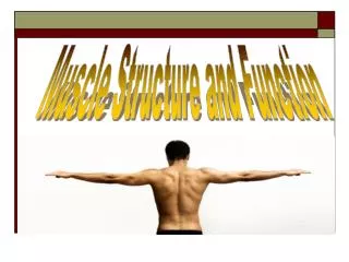
Muscle Structure
Muscle Structure. Animal Growth and Development. Types of Muscle. Skeletal Cardiac Smooth Classification Striated Non-striated Voluntary Involuntary. Skeletal structure. Based on size, shape and location Origin- where muscles originate on one side of the joint

Muscle Structure
E N D
Presentation Transcript
Muscle Structure Animal Growth and Development
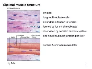
Types of Muscle • Skeletal • Cardiac • Smooth • Classification • Striated • Non-striated • Voluntary • Involuntary
Skeletal structure • Based on size, shape and location • Origin- where muscles originate on one side of the joint • Insertion- where muscles terminate on the other side of the joint • Tendon – point of attachment at either insertion or origin • Fascia- a thin sheet of connective tissue at attachment
Organization • Epimysium • Perimysium • Endomysium Sarcolemma Muscles are multinucleated
Muscle Cytoskeleton • Microfilaments • Intermediate filaments • Microtubules • Myofibrils are microfilamentous organelles of muscle fibers • Sarcomere – smallest contractile unit of muscle
Myofibrils • Made up of thick and thin filaments called myosin and actin • Z-line – outer demarcation of the sarcomere • Made up predominantly of alpha-actinin • A- band • H – Zone • I - band
Myosin • Occupies 80-87% of the total volume of the muscle fiber • Contains both heavy and light chain molecules (heavy and light meromyosin) • Positioned in the center of the sarcomere • Rachet effect – acts in contraction to make the sarcomere shorten because it pulls on actin filaments, thus shortens one Z-line to another
Actin • Second most abundant in muscle fibers and makes up almost 20% of the total protein • Filamentous actin (F-actin) is predominant • G-actin or globular is smaller yet is found to aid in the development of F-actin • Numerous G-actins develop one F-actin
Regulatory proteins • Tropomyosin – another filamentous protein of the thin filament • Troponin – also is a filamentous protein that surrounds actin and holds the actin strands together • Troponin is involved in calcium binding upon release of or increased calcium levels in the muscle to initiate contraction
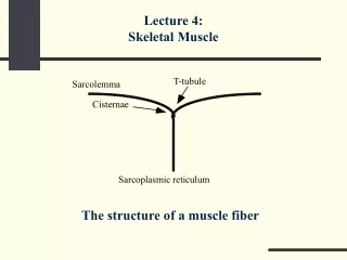
Additional Components of Muscle Fibers • Sarcoplasmic reticulum – membranous system of tubules that forms a network around each myofibril • T – tubules – (transverse tubules) surround myofibrils at the A-I band jct. • Figure 2.34 • In fact, the system between two T-tubules is called the Sarcoplasmic reticulum
Additional Components of Muscle Fibers • Sarcoplasmic reticulum consists of two structures • Terminal cisternae • Fenestrated collar • These act as Ca storage reservoirs • The structure formed from the two terminal cisternae and on each side of the T-tubule is called a triad. Note: there is no physical contact between the T-tubule and the terminal cisternae • Communication occurs via electrical messages
Additional Components of Muscle Fibers • The electrical system • Dihydropyridine and ryanodine recptors • When an electrical stimuli occurs at these receptors, the SR channel changes conformation and allows Ca into the cytoplasm • Calsequestrin is the part of the fiber/cell (protein) that binds calcium in the SR • Nucleus – muscle fibers are multinucleated • Mitochondria- most of the energy is made • Sarcoplasm – cellular cytoplasm of the muscle cell
Cardiac Muscle • Cardiocytes –cardiac muscle fibers • Striated and involuntary (uniform contractions) • Communication of these type contractions occurs at connections called intercalated discs • Cardiac myofibrils are attached directly to the intercalated discs and allow the heart to act in syncytium or as a group
Cardiac muscle • Cardiac thin filaments are more variable in length as compared to skeletal muscle fibers • Cardiac muscle must contract even under extreme conditions • Because of the increased variability, this allows more overlap for contraction during stressed times for more uniform, consistent contraction • Cardiac muscle fibers are more branched and have more mitochondria than skeletal muscle
Smooth muscle • Seen as sheet-like muscle masses • Is very elastic and pliable • One example is around the uterus where it has to stretch • It thickens during pregnancy and becomes more thin while the female is open • Smooth muscle is more triangular and only has one nucleus per cell • Thick and thin filaments are not symmetrically arranged like striated muscle
Smooth muscle • As compared to skeletal muscle, smooth muscle has dense bodies that are analogous to Z-lines • Actin molecules are attached to dense bodies • Thick filaments are likewise positioned throughout the cells that overlap the thin filaments • As a result, when contraction occurs, forces react in varying directions
Myogenesis • Origin of muscle which begins at fertilization • Develops from mesodermal somites • DNA responsibility • Muscle is continually modified throughout growth and development • Mitosis – increase in cell numbers or cell division • During mitosis, the entire genome or DNA has to be replicated • Figure 4.4
Muscle cell cycle • Four distinct phases • Gap 1 (G1) all cells enter the cell cycle from this phase • Most variable phase in length • Respond to cues from the environment • Synthetic or S-phase is the time where the cell dedicates to DNA replications or synthesis • Third phase is called G2, and follows the S-phase
Muscle cell cycle • G2 phase represents when the intracellular architecture remodels itself to accommodate physical division of the cell mass in mitosis or M-phase • M-phase is the shortest phase • After this, it re-enters the G1 phase • Growth factors are often small proteins that influence the environment that initiate cell growth
Muscle cell cycle • These small proteins bind to receptors • Also, steroids can bind and create an environment for growth • Ex. IGF-1, testosterone, etc. • Another alternative for cells in the G1 phase is to exist in a protracted G1 phase or G0 phase. • Cells are capable of remaining dormant, which can re-enter the proliferative cycle without dividing • These cells in muscle are called satellite cells
Muscle cell determination • Determination • Cells must migrate from the somites (matured mesodermal cells (Fig. 3.1 and 3.2) • Cells that are determined to develop into muscle are termed as myoblasts (precursors for muscle fibers) Fig. 4.4 • Muscle regulatory factor gene (MRFs) • These trigger the expression of one or more genes for myogenesis
Muscle cell determination • Transcription factors include MRFs and are responsible for turning on transcription of other genes located in the nucleus and contain helix-loop-helix motif • When two of these proteins combine they bind to the regulatory region and cause the gene to be expressed or repressed • Thus, MRFs are control point for myogenesis
Muscle cell determination • Helix-loop-helix proteins include: • Myogenin • MRF-4 • Myo-D • Myf-5 • Fig. 4.5 MRFs action
Basic Genetics and CytogeneticsTwo Basic Concepts • DNA- Polymer of Nucleotide Bases • Complementary Base Pairs • 1 Gene – 1 Protein (Gene determining the Phenotype) DNA is the Genetic Code for Protein Synthesis • Gene is the Unit of inheritance (Inherited from both Parents) Complementary Base Pares – DNA Duplication and Gamete production.
Cell Division – Mitosis - MeiosisDNA- Contain the Genes – The unit of Inheritance DNA- Complementary Bases
Chromosomes- Chromatin and DNA DNA – Genetic Code for Protein SynthesisDNA – Polymer of Nucleotide Bases Nucleotide Sequence –Amino acid Sequence of the Protein (Shape and function)
I - DNA is the Genetic Code for Protein SynthesisProteins can be Enzymes, Hormones, Transcription Factors, Structural, etc.Nucleotide Sequence –Amino acid Sequence of the ProteinShape Determine the function/effect)
Differentiation and Fusion • Replication competent – capable of progressing the cell cycle and giving rise to additional myoblasts even though determined muscle cells & myoblasts have express muscle regulatory factor genes • Myoblasts do not contract because they do not contain contractile proteins yet • Before this, cells must receive signals that induce them to differentiate
The “Differentiating” process • Cells stop dividing • Cells begin to align with one another • Cell membranes begin to fuse together to form an immature muscle fiber (myotube) • Muscle specific genes are regulated to initiate • Genes for muscle creatine kinase and acetylcholine receptor subunit, myosin and actin are triggered
Fusion • The exact process is still cloudy • Yet, we know that small tubules or attachments are apparent and dense structures beneath membranes are generated to form tight junctions between myoblasts • Finally, two bilipid membranes become one and dissolve in the cytoplasm of the newly formed multinucleated cell. Note: Ca ions play a significant role in this process
Maturation • Once myoblasts differentiate and fuse to form myotubes, resulting cells do not continue to express a given set of tenes • The new muscle fibers change following fusion • This process is called maturation
Morphological Aspects of Myogenesis • Once the somite has reached the two layered, dermomyotome stage cells begin to accumulate ventrally to form the myotome. • Myotome – a compartment of the somite • This is believed to involve migration of cells from the dorsomedial ridge and ventrolateral part of the dermomyotome to below it creating the myotome
Morphological Aspects of Myogenesis • Recall that skeletal muscle originates from somites which is developed from the mesoderm • Myogenesis appears to involve migration from the dorsomedial ridge and ventrolateral spects of the dermomyotome to immediately below the dermomyotome, thus creating a myotome Fig 4.8
Morphological Aspects of Myogenesis • Muscle does not just appear during prenatal development • It results from biphasic processes • Occurs from two populations of muscle fiber myoblasts • During myoblast differentiation, myoblasts cluster align in a vast network of connective tissue and fuse to form an immature muscle fiber referred to as a myotube
Morphological Aspects of Myogenesis • Primary myotubes are the first to develop • These primary myotubes are the structure for which others attach and complete the final fiber structure - Figure 4.11 • The myotube formation can be referred to as the second phase of myogenesis (fetal myoblasts) whereas the first phase of primary myotubes are referred to as embryonic myoblasts
Morphological Aspects of Myogenesis • Upon receiving signals to differentiate, the secondary myotube use the primary myotubes as a template for alignment and organization • This alignment of fetal with embryonic myotubes align closely and facilitates the fusion process • After contraction the primary fibers remain intact and constant whereas the secondary muscle fibers or myotubes are splintered away • This process of producing primary myotubes is self-governing in nature
Morphological Aspects of Myogenesis • Depending on the species, muscle development occurs during the first 2/3 of prenatal development • The total # of myofibers is considered established by 90 days post-conception with pigs Fig. 4.12 - K State gestation study • As muscle growth increases, muscle fiber diameter increases
Myofibrillogenesis • The primary f(x)n of muscle is to contract • This is done by the addition of myuofibrils to each cell during development • Again, skeletal muscle is highly organized and contains many proteins • Stress fibers are those that are bundles of myofilaments which have contractile possibilities
Myofibrillogenesis • The stress cells are located at the periphery and interact with membrane bound proteins called extracellular and cell-adhesion molecules • Integrins (extracellular adhesion molecules) are proteins that interact with extracellular proteins of connective tissues • Two families that are responsible for holding cells close in contact are: adherins and NCAM’s • Development of stress fibers to the cell membrane represents a template for the development of nascent myofibrils (those initially responsible for developing muscle cells)
Muscle Growth • Hypertrophy vs Hyperplasia Fig. 5.1 • Muscle fibers are oriented in an oblique fashion rather than parallel to the long axis • Muscle fiber # is best assessed when muscles contain more fibers that are arranged in the long axis orientation • Individual muscle fibers do not necessarily extend the entire length of the muscle
Muscle Growth • Mean packing density = # of fibers per unit of a cross-sectional area • Again, minimal increases in #’s of fibers occur during postnatal development • The occurrence when reported that #’s increased was due to a muscle fiber lengthening process • This may occur during muscle regeneration following an injury or some type of death to the myofiber
Muscle Growth • Satellite cells of the muscle fiber that are located between the sarcolemma and basal lamina are activated to to proliferate and ultimately fuse with other satellite cells to form new fibers
Factors affecting muscle fiber numbers • Animal variation • Varies from animal to animal and from muscle to muscle • Muscle • The primary diff. in the size of muscle is the number of muscle fibers contained within each • This may range from a few thousand to billions depending on the muscle • What would be one of the largest muscles????
Factors affecting muscle fiber numbers • Species • Excess muscle growth both #’s and size (both length and diameter) contribute to the size of muscles in various species • Also, size of muscle fibers and thus muscles contribute to the variation between species • this would include mature cattle versus sheep, etc. • Porcine semitendinosus has roughly 1/3 of the # of fibers as does the beef semitendinosus muscle
Factors affecting muscle fiber numbers • Nutrition • Nutrition has a greater impact on muscle at specific stages of growth thus altering muscle development at various stages of growth is imperative • Younger animals require more protein for muscle growth than older animals • Also, in young animals, 600 lb. calves require more protein than a 900 lb. calf and those require more protein than those at 1000 lbs.
Factors affecting muscle fiber numbers • Swine – litter bearing species has to prioritize across the embryos and fetuses; thus, variations of muscle development among littermates occur often • Genetic differences may occur among siblings • Ex. Runts – those that weigh less than 2/3 of the mean wt. of a given litter
Factors affecting muscle fiber numbers • Runts ex. • If they survive, they will usually enter the fattening phase earlier (earlier maturity pattern) and not be as heavy muscled. • This may be due to undernourishment or simply lesser development prenatally • Yet, if adequate nutrition was available during fetal muscle development, then numbers of muscle fibers are not affecting during nutrition depravation during a postnatal state.
Factors affecting muscle fiber numbers • Age – the # of muscle fibers that an animal develops is fixed at birth because hyperplasia occurs “in ovo” or “in utero” • Muscle fibers continue to die and regenerate with age • Yet, with age muscle fiber number does not change but when tissue mass is no longer maintained then fibers are lost