Download
1 / 10
120 likes | 275 Views
Thoracic Cavity and Respiration. Alveoli Respiratory Tree Lungs Diaphragm Intercostal Muscles. Alveoli. Key to lung function Where O2 enters blood, CO2 leaves blood Every alveoli is capillary covered sac Lining of sac is squamous epithelium

E N D
Thoracic Cavity and Respiration • Alveoli • Respiratory Tree • Lungs • Diaphragm • Intercostal Muscles Larry M. Frolich, Human Anatomy, Respiratory Function
Alveoli • Key to lung function • Where O2 enters blood, CO2 leaves blood • Every alveoli is capillary covered sac • Lining of sac is squamous epithelium • Also cuboidal epithelial cells that secret surfactant (keeps surfaces from sticking) and cilia (to move mucous and particles up respiratory tree Larry M. Frolich, Human Anatomy, Respiratory Function
Respiratory Tree (how does air get to alveoli?) • Branching pattern • Trachea • Primary bronchi (left, right) • Secondary bronchi (each lobe) • Tertiary bronchi, bronchioles • Alveoli • Vessels accompany respiratory tree to alveoli Larry M. Frolich, Human Anatomy, Respiratory Function
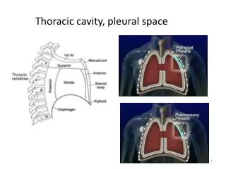
LUNGS • Located in Pleural Cavities • Lateral to Mediastinum • Location • Apex posterior to clavicle • Base lays on Diaphragm • Costal Surface = Ant, Lat, Post surfaces contact wall of chest cavity • Root - all vessels, nerves enter each lung • 2 Pulmonary Veins = carries O2 blood from each lung to heart • 1 Pulmonary Artery = carries De-O2 blood to each lung • Primary Bronchi • Nerves • –Lymph Vessels Pg 618 Larry M. Frolich, Human Anatomy, Respiratory Function
LOBES OF LUNGS • Left Lung = 2 lobes • Upper • Lower • Oblique Fissure • Cardiac Notch • Right Lung = 3 lobes • Upper • Middle • Lower • Oblique fissure • Horizontal fissure Pg 622 Larry M. Frolich, Human Anatomy, Respiratory Function
Specific Location of Lungs • Right Lung • 1” above Rib 1 • Crosses Costal Cartilage 6 • Midclavicular at Rib 6 • Midaxillary at Rib 8 • Vertebral Border at Rib 10 • Inferior border 2 rib widths above diaphragm • Left Lung • 1” above Rib 1 • Deep to Manubroclavicular joint • Midsternally to Rib 4 • Jogs to left, continues to Rib 6 • Midaxillary Rib 8 • Vertebral Border at Rib 10 Larry M. Frolich, Human Anatomy, Respiratory Function
Action of the Diaphragm • Primary muscle of respiration (involuntary) • Contraction during inspiration • Increases volume of thoracic cavity • Decreases pressure of thoracic cavity • Air moves into lungs (highlow pressure) • Forced contraction (voluntary) • Used for defecation, urination, labor • Increases pressure in abdominal cavity • Pushes on abdominal organs to move contents out Larry M. Frolich, Human Anatomy, Respiratory Function
Anatomy of the Diaphragm • Skeletal Muscle • Dome-shaped (relaxed) • Divides thoracic & abdominal cavities • Attachments • Origin: Inferior Internal rib cage • Insertion: Central tendon • Innervated by right + left PHRENIC Nerves Superior View Pg 288 Larry M. Frolich, Human Anatomy, Respiratory Function
INTERCOSTAL MUSCLES • Lift ribs to expand chest cavity for inspiration • Three layers Larry M. Frolich, Human Anatomy, Respiratory Function