Download
1 / 28
290 likes | 706 Views
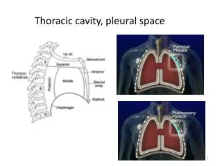
Thoracic cavity, pleural space. Conditions requiring chest drainage_1. Air between the pleurae is a pneumothorax Occurs when there is an opening on the surface of the lung or in the airways, in the chest wall — or both

E N D
Conditions requiring chest drainage_1 Air between the pleurae is a pneumothorax • Occurs when there is an opening on the surface of the lung or in the airways, in the chest wall — or both • The opening allows air to enter the pleural space between the pleurae, creating an actual space
Conditions requiring chest drainage_2 Blood in the pleural space is a hemothorax Lateral decubitus X-Ray
Conditions requiring chest drainage_3 • pleural effusion • Transudate • Exudate • Empyema:
open vs closed pneumothorax Open pneumo • Opening in the chest wall (with or without lung puncture) Closed Pneumo • Chest wall is intact • Rupture of the lung and visceral pleura (or airway) allows air into the pleural space Photo courtesy trauma.org
tension pneumothorax • Tension pneumothorax occurs when a closed pneumothorax creates positive pressure in the pleural space that continues to build • That pressure is then transmitted to the mediastinum (heart and great vessels)
mediastinal shift from a tension pneumothorax • Mediastinal shift occurs when the pressure gets so high that it pushes the heart and great vessels into the unaffected side of the chest • These structures are compressed from external pressure and cannot expand to accept blood flow Mediastinal shift
Clinical Manifestations of a collapsed lung • SOB • Chest Pain • Cough • Absent or decreased breath sounds on affected side • Shallow Respirations • Asymmetrical chest movement • Decreased O2 saturation
Treatment for pleural conditions 1. Remove fluid & air as promptly as possible 2. Prevent drained air & fluid from returning to the pleural space • Restore negative pressure in the pleural space to re-expand the lung
Remove Fluid &/or Air: chest tube insertion • Chest tube tray with an appropriate size tube • Surgical prep, sutures, sterile gloves • Lidocaine, needles, syringes, alcohol preps • Vaseline gauze, 4x4s & tape • CDU = Chest drainage unit • Suction and sterile water
RN Role • Educate patient and family • Administer pain meds • Set up chest drainage unit • Obtain consent • Assists with insertion PRN • Verify occlusive dressing is intact • Tape all connections from CT to drainage system to prevent air leaks • Assess the patient and document appropriately
2. Prevent air & fluid from returning to the pleural space Chest tube is attached to a drainage device • Allows air and fluid to leave the chest • Contains a one-way valve to prevent air & fluid returning to the chest • Designed so that the device is below the level of the chest tube for gravity drainage
3. Restore negative pressure in the pleural space Tube to vacuum source Tube open to atmosphere vents air Tube from patient Straw under 20 cmH2O Fluid drainage Suction control 2cm fluid water seal Collection bottle
Restore negative pressure in the pleural space The depth of the water in the suction bottle determines the amount of negative pressure that can be transmitted to the chest, NOT the reading on the vacuum regulator
How a chest drainage system works: summary • Expiratory positive pressure from the patient helps push air and fluid out of the chest (cough, Valsalva) • Gravityhelps fluid drainage as long as the chest drainage system is below the level of the chest • Suction can improve the speed at which air and fluid are pulled from the chest
Collection Chamber • This chamber allows monitoring of volume, rate and nature of the drainage • Measure output per hospital policy • Most systems are considered “full” at 2500ccs
Water Seal Chamber • Water creates a one-way valve that prevents air or fluid from returning to the patient’s chest • Monitor this chamber for: • air leaks (bubbling) • tidaling (fluctuations in fluid level) • increased negative pressure
Suction Control Chamber • regulates the suction level acceptable for thoracic drainage • Suction increases drainage rate • Suction is controlled by water level • Regulate wall suction until gentle bubbles appear
Monitoring air leak • Water seal is a window into the pleural space • Not only for pressure • If air is leaving the chest, bubbling will be seen here • Air leak meter (1-5) provides a way to “measure” the leak and monitor over time – getting better or worse?
Air Leaks • Continuous bubbling initially - OK • Bubbling when pt coughs or exhales. • How to troubleshoot: • Crepitus (subcutaneous emphysema)
Tubing from chest drainage system • Make sure connections are tight and taped • No Dependant loops • Milking or Stripping- only done if clot is suspected • Controversial : may cause damage to lung tissue as increased negative pressure is exerted
Transporting a patient with a chest tube • Keep the drainage system lower than the patients chest • May open suction end to air which equals a water seal • Mayo clamps (rubber tipped hemostats) should be kept at the bedside
Then: Assess the CDU • Check the dressing • Check tubing - dependent loops • Check drainage in tubing & collection chamber • Check water seal chamber • Bubbling • tidaling • Check level of water • Water seal chamber • suction control chamber • Check tubing CDU to wall suction: open?
Accidental disconnection of tube and drainage system • Reconnect ASAP or • Place end of tube in a sterile water bottle until new system arrives • Monitor patient for s/s of resp distress • Notify physician
Accidental DC of Chest Tube • Seal off insertion site – dry, sterile dressing or, petroleum gauze dressing • secure on 3 sides • Notify physician • Assess patient prepare to assist with reinsertion • Watch for tension pneumothorax
Termination of Chest Tube • Assess for signs of re-expansion • Minimal drainage • Minimal bubbling / fluctuations in water seal chamber • Chest x-ray shows re-expansion • MD may leave to gravity 24°
Termination of Chest Tube • Explain procedure to patient • Equipment • Suture removal kit, gloves, Vaseline gauze, • 4x4s, tape, towels • Tube should be pulled at the end of full inspiration. • Some physicians prefer coughing or holding breath to increase intrathoracic pressure • Occlusive dressing