THORACIC WALL
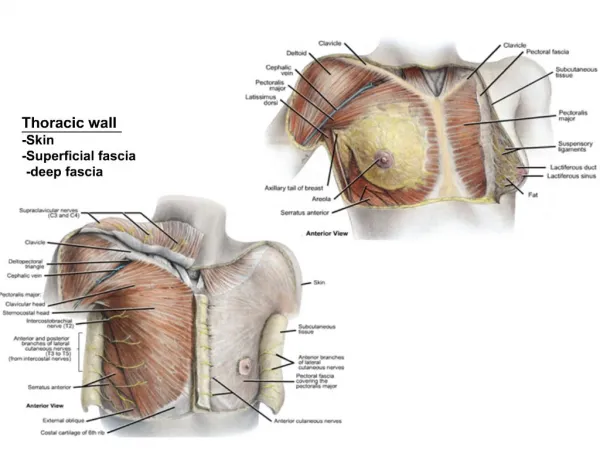
THORACIC WALL. MUSCLES, MAMMARY GLANDS, CROSS-SECTION. THORACIC WALL MUSCLES. Surface Muscles (Anterior). Platysma Pectoralis major Pectoralis minor Subclavius Serratus anterior Refer in syllabus: Table 1, pp 47-49 Figure 13, p 50. Surface Muscles (Posterior). Latissimus dorsi


THORACIC WALL
E N D
Presentation Transcript
THORACIC WALL MUSCLES, MAMMARY GLANDS, CROSS-SECTION
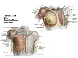
Surface Muscles (Anterior) • Platysma • Pectoralis major • Pectoralis minor • Subclavius • Serratus anterior • Refer in syllabus: Table 1, pp 47-49 Figure 13, p 50
Surface Muscles (Posterior) • Latissimus dorsi • Trapezius • Rhomboideus major • Rhomboideus minor • Refer in syllabus: Table I; pp 47-9
Deltopectoral Triangle • Boundaries: Anterior border of the deltoid. Superior border of the pectoralis major. Middle third of the clavicle.
Deltopectoral Triangle • Contents: Cephalic vein. Deltopectoral lymph nodes. Deltoid branch of the thoracoacromial artery.
Clavipectoral Fascia • Invests subclavius and pectoralis minor. • Attached to clavicle and anterior thoracic wall. • Pierced by: Cephalic vein Thoracoacromial artery Lateral pectoral nerve • Become suspensory ligament of the axilla.
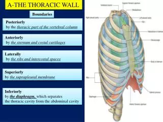
THORACIC WALL STRUCTURE (x.s.) Refer to Figure 15, p. 60 in syllabus as well as background material on pp 58-59.
Mammary Glands • Modified sweat glands • 15-20 lobes • Lobes separated by fibrous septa • Lactiferous duct (1 per lobe) • Lactiferous sinus (ampulla): Dilation as duct enters nipple
Mammary Glands • Fibrous tela subcutanea: Connective tissue layer surrounding the entire gland. • Fatty tela subcutanea: Adipose tissue deep to fibrous layer. • Suspensory ligament of Cooper: Bundles of collagen fibers in dermis and hypodermis.
Mammary Glands • Breast extends from 2nd-3rd rib superiorly to 6th-7th costal cartilage inferiorly. • Extends from lateral border of sternum to beyond the anterior axillary fold.
Mammary Glands • Retromammary space: Space between the gland and the pectoralis major muscle. • Sinus mammarumis: Space between the two glands.
Mammary Gland Arteries • Anterior perforating arteries: From internal thoracic artery To medial part of gland • Medial mammary rami: From 2nd - 4th anterior perforating arteries To deep medial part of gland
Mammary Gland Arteries • Lateral mammary artery: From lateral thoracic artery. To inferior part of gland. • Lateral mammary rami: From lateral cutaneous branches of intercostal arteries. To lateral part of the gland.
Mammary Gland Veins • Superficial and deep venous plexuses drain into internal thoracic, lateral thoracic, and intercostal veins.
Mammary Gland Nerves • Lateral mammary nerve: T2-T6 • Anterior branch of lateral cutaneous branch of intercostal nerves. • Medial mammary nerve: T2-T6 • Lateral branch of anterior cutaneous branch of intercostal nerves.
Mammary Gland Lymphatics • Perilobular and interlobular lymphatic vessels: Into: • Subareolar plexus: Into: • Lateral lymphatic trunk: From lateral and superior gland • Medial lymphatic trunk: From medial and inferior gland
Mammary Gland Lymphatics • Lateral lymphatic trunk and Medial lymphatic trunk: Into: • Pectoral group of axillary lymph nodes: Into: • Subclavian lymphatic channels
Mammary Gland Lymphatics • Accessory lymphatic drainage: Periphery of gland drains into apical group of axillary nodes and follows thoracoacromial trunk. Circumareolar channels drain into sternal chain.
Lymphatic Drainage • Subareolar plexus of nodes • Axillary lymph nodes: Receive from: Superficial tissues, skin, breast, extrinsic limb muscles. Include: Pectoral group Lateral group Apical group Subscapular group
Lymphatic Drainage • Infraclavicular nodes • Parasternal nodes • Abdominal nodes
Inspiration: “Bucket Handle” • Involves contraction of intercostal muscles • Results in raising of ribs • Results in an increase in the lateral dimensions of the thoracic cage.
Inspiration: “Pump Handle” • Results from raising of sternum • Results in increase in anteroposterior dimensions of thoracic cage
Abdominal Breathing • Results from lowering of diaphragm: Phrenic nerve • Necessary when: Infant Costal cartilages are calcified
Expiration • Mostly passive