Download
1 / 70
700 likes | 879 Views
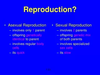
Human. Reproduction. Sexual Reproduction in Human. testis. ovary. meiosis. meiosis. sperms. eggs (ova). fertilization. zygote. embryo. foetus. baby. Male Reproductive System. vas deferens (sperm duct). epididymis. Click here. testis. Testes.

E N D
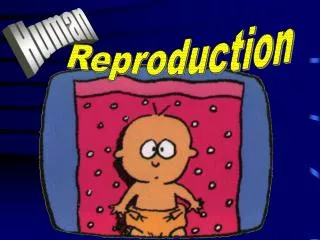
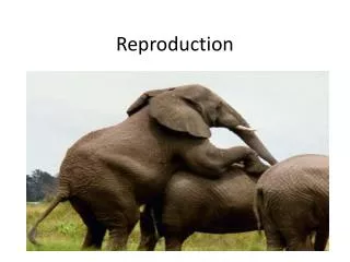
Human Reproduction
Sexual Reproduction in Human testis ovary meiosis meiosis sperms eggs (ova) fertilization zygote embryo foetus baby
Male Reproductive System vas deferens (sperm duct) epididymis Click here testis
Testes • For production of male gametes (sperms) • For production of male sex hormones Click here
Epididymis • For temporarily storage of sperms • During copulation, muscles of epididymis contract to release sperms Click here
Testis and epididymis epididymis vas deferens (sperm duct) sperm tubules
Male Reproductive System seminal vesicle secrete seminal fluid prostate gland Cowper’s gland
Functions of Seminal Fluid • To provide a medium for the sperms to swim • To activate and nourish the sperms • To neutralize the acidity in the female reproductive tract Seminal Fluid Semen Sperms +
Male Reproductive System urethra penis
Penis • Erected during copulation for insertion into vagina • Dilation of arterioles causes the erectile tissue of penis become turgid • Muscles of epididymis contract • Semen is squeezed from the penis to the top of vagina • Ejaculation
The human penis • Is composed of three cylinders of spongy erectile tissue • During sexual arousal • The erectile tissue fills with blood from the arteries, causing an erection
Female Reproductive System oviduct ovary uterus cervix vagina Video of fertilization Click here
Functions of Ovary • For production of female gametes (ova/eggs) • For production of female sex hormones Click here
Oviduct • Carries the ovum forward by • the beating action of the cilia on its inner surface • the contraction of muscles of oviduct Click here
Ovulation • The release of an ovum from an ovary Video of ovulation Video of ovulation
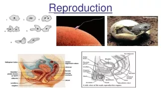
Structure of human gametes Sperm (male) Ovum (female) Size • Larger (0.1mm) • with large amount of food for early development of embryo • Smaller (0.05mm) • to reduce energy for movement • to allow faster movement Position of nucleus In the head Towards centre Very active, moves by its tail Not able to move by itself Movement
Structure of human gametes II 0.05 mm 0.1 mm Sperm (male) Ovum (female) Tadpole-like, with a head and a tail Spherical Shape plasma membrane clear membrane tail head nucleus cytoplasm nucleus follicle cells
Oogenesis differs from spermatogenesis In three major ways…….……
First, during the meiotic divisions of oogenesis Cytokinesis is unequal, with almost all the cytoplasm monopolized by a single daughter cell, the secondary oocyte • Second, sperm are produced continuously throughout a male’s life • Which is not the case in oogenesis • Third, oogenesis has long “resting” periods / arrested at certain stages • While spermatogenesis produces sperm in uninterrupted sequence
Transfer of semen and fertilization when sexually stimulated, arterioles dilate and erectile tissue filled with blood, penis becomes hard and erect vas deferens testis soft penis not sexually stimulated
Transfer of semen and fertilization During copulation hard and erect penis inserts into vagina muscles of epididymis contract semen containing millions of sperms is ejaculated into the vagina
Menstrual Cycle • Once in about 28 days • The uterine lining becomes thickened 14 days after ovulation to prepare for the fertilized ovum to implant in it
Video on change in uterine lining Menstrual Cycle • Day 6 - 14 • Lining becomes thicker with increased blood supply • Day 14 : ovulation • Day 14 - 28 • Lining remains thick to ready for implantation of fertilized ovum • Day 28 • No implantation of fertilization ovum • Uterine lining breaks down; menstruation starts • Day 1 - 5 • Menstruation starts • Uterine lining decreases in thickness to a minimum
Transfer of semen and fertilization site of fertilization • sperms meet the ovum in oviduct • sperms swim up the uterus • sperms swim through the cervix • sperms ejaculated into the vagina during copulation
one sperm penetrates the egg membrane site of fertilization clear membrane hardens; no other sperms can enter female nucleus male nucleus fuses with the female nucleus
20.7 The development of the human embryo • zygote undergoes repeated mitotic cell divisions • ovum fertilized by a sperm in oviduct pregnancy begins • ovum is released from ovary • embryo implants into the uterine wall Stages leading to implantation
Events Happened after Fertilization • Ovum is fertilized at the oviduct • Fertilized ovum is carried to the uterus by • the beating cilia on the inner wall of oviduct • the contraction of muscles of oviduct • After reaching the uterus, the fertilized ovum fixed firmly onto the thick uterine wall Implantation
Development of Human Foetus uterus foetus placenta umbilical cord amnion amniotic fluid Video on foetus development Click here
Functions of the Uterus • During embryo development • Protect the embryo • Provide a constant environment for the embryo to develop • Allow placenta to attach on • During birth of baby • Push the baby out by muscular contraction Click here
Functions of the Amniotic Fluid • To keep the foetus moist to prevent dessication • As a water cushion to • support the foetus • allow it to move freely • absorb shock • protect the foetus from mechanical injuries • To reduce temperature fluctuation • To lubricate the vagina during birth Click here
The Placenta oxygenated blood from mother’s artery deoxygenated blood to mother’s vein villus umbilical vein umbilical artery
After implantation uterus mother’s blood space close contact placenta blood capillaries of embryo • temporary disc-shaped organ embryonic villus which grows into the uterine wall umbilical cord umbilical vein umbilical artery • allows exchange of materials between embryo & mother • connected to embryo via umbilical cord embryo
After implantation uterus amniotic fluid • protects embryo against shock • keeps a constant environment • prevents desiccation • allows embryo to move easily as embryo develops, amnion is formed secretes amniotic fluid embryo
Functions of the Placenta • As a place of exchange of materials between mother and the foetus • For secreting hormones
Adaptations of the Placenta • Finger-like villi • to increase the surface area for efficient diffusion • Maternal blood and foetal blood flows in opposite direction • to speed up diffusion of materials between them • Maternal blood capillaries and foetal blood capillaries are separated by thin membrane • to shorten the distance of diffusion of materials
Adaptations of the Placenta • Maternal blood is separated from foetal blood by capillary wall • to prevent high pressure of maternal blood to break the delicate foetal blood vessels • to prevent harmful substances to enter the foetus • to prevent clotting of maternal and foetal blood if their blood groups are incompatible Click here
How is the baby born? A few weeks before birth foetus changes its position, so that the head is downward As birth approaches uterine muscles start to make rhythmic contractions • labour begins
abdominal muscles How is the baby born? uterine wall Birth process (3 stages) amnion • The dilation stage • cervix dilates • uterine & abdominal muscles contract • amnion breaks • amniotic fluid comes out amnion fluid
How is the baby born? Birth process (3 stages) • The expulsion stage • muscular contractions push the foetus out (usually head-first) • umbilical cord is tied and cut
How is the baby born? umbilical cord uterus Birth process (3 stages) • The placental stage • placenta is pushed out of the body placenta (partially detached)
The Birth Process • Onset of labour • Uterine muscles begin to make rhythmic contractions • Contractions gradually become stronger and closer
The Birth Process • Uterine contractions causes amnion to break and amniotic fluid to escape out of the vagina • Muscular contractions push the foetus head first through the vagina, and the umbilical cord is cut and tied • Further contractions push the placenta out of the body • “After birth” • Dilation of cervix allow the head of the foetus to pass through
How is the baby born? After birth • baby begins to breathe a few seconds after being born navel • umbilical cord shrivels and falls away • a scar (navel) is left
Could you survive after birth without the care of your parents?
Parental Care • To increase the chance of survival of the young • Mother feeds milk to the baby • Milk provides the babies with a balanced diet • It also contains antibodies which defend the babies against infection