Reabsorption and Secretion
Reabsorption and Secretion. Hydrogen Ion Secretion Are generated by dissociation of carbonic acid by enzyme carbonic anhydrase Secretion is associated with reabsorption of sodium Secreted by sodium-linked countertransport In exchange for Na + in tubular fluid


Reabsorption and Secretion
E N D
Presentation Transcript
Reabsorption and Secretion • Hydrogen Ion Secretion • Are generated by dissociation of carbonic acid by enzyme carbonic anhydrase • Secretion is associated with reabsorption of sodium • Secreted by sodium-linked countertransport • In exchange for Na+ in tubular fluid • Bicarbonate ions diffuse into bloodstream • Buffer changes in plasma pH
Reabsorption and Secretion • Hydrogen Ion Secretion • Acidifies tubular fluid • Elevates blood pH • Accelerates when blood pH falls
Reabsorption and Secretion Figure 24–14c Tubular Secretion and Solute Reabsorption at the DCT.
Reabsorption and Secretion • Acidosis • Lactic acidosis • Develops after exhaustive muscle activity • Ketoacidosis • Develops in starvation or diabetes mellitus
Reabsorption and Secretion • Control of Blood pH • By H+ removal and bicarbonate production at kidneys • Is important to homeostasis
Reabsorption and Secretion • Alkalosis • Abnormally high blood pH • Can be caused by prolonged aldosterone stimulation • Which stimulates secretion
Reabsorption and Secretion • Response to Acidosis • PCT and DCT deaminate amino acids • Ties up H+ • Yields ammonium ions (NH4+) and bicarbonate ions (HCO3-) • Ammonium ions are pumped into tubular fluid • Bicarbonate ions enter bloodstream through peritubular fluid
Reabsorption and Secretion • Benefits of Tubular Deamination • Provides carbon chains for catabolism • Generates bicarbonate ions to buffer plasma
Reabsorption and Secretion • Reabsorption and Secretion along the Collecting System • Collecting ducts • Receive tubular fluid from nephrons • Carry it toward renal sinus
Reabsorption and Secretion • Regulating Water and Solute Loss in the Collecting System • By aldosterone • Controls sodium ion pumps • Actions are opposed by natriuretic peptides • By ADH • Controls permeability to water • Is suppressed by natriuretic peptides
Reabsorption and Secretion • Reabsorption in the Collecting System • Sodium ion reabsorption • Bicarbonate reabsorption • Urea reabsorption
Reabsorption and Secretion • Secretion in the Collecting System • Of hydrogen or bicarbonate ions • Controls body fluid pH
Reabsorption and Secretion • Low pH in Peritubular Fluid • Carrier proteins • Pump H+ into tubular fluid • Reabsorb bicarbonate ions
Reabsorption and Secretion • High pH in Peritubular Fluid • Collecting system • Secretes bicarbonate ions • Pumps H+ into peritubular fluid
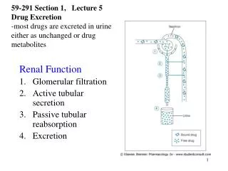
Reabsorption and Secretion • The Control of Urine Volume and Osmotic Concentration • Through control of water reabsorption • Water is reabsorbed by osmosis in • Proximal convoluted tubule • Descending limb of nephron loop
Reabsorption and Secretion • Water Reabsorption • Occurs when osmotic concentration of peritubular fluid exceeds that of tubular fluid • 1–2% of water in original filtrate is recovered • During sodium ion reabsorption • In distal convoluted tubule and collecting system
Reabsorption and Secretion • Obligatory Water Reabsorption • Is water movement that cannot be prevented • Usually recovers 85% of filtrate produced
Reabsorption and Secretion • Facultative Water Reabsorption • Controls volume of water reabsorbed along DCT and collecting system • 15% of filtrate volume (27 liters/day) • Segments are relatively impermeable to water • Except in presence of ADH
Reabsorption and Secretion • ADH • Hormone that causes special water channels to appear in apical cell membranes • Increases rate of osmotic water movement • Higher levels of ADH increase • Number of water channels • Water permeability of DCT and collecting system
Reabsorption and Secretion • Osmotic Concentration • Of tubular fluid arriving at DCT • 100 mOsm/L • In the presence of ADH (in cortex) • 300 mOsm/L • In minor calyx • 1200 mOsml/L
Reabsorption and Secretion • Without ADH • Water is not reabsorbed • All fluid reaching DCT is lost in urine • Producing large amounts of dilute urine
Reabsorption and Secretion Figure 24–15 The Effects of ADH on the DCT and Collecting Duct.
Reabsorption and Secretion • The Hypothalamus • Continuously secretes low levels of ADH • DCT and collecting system are always permeable to water • At normal ADH levels • Collecting system reabsorbs 16.8 liters/day (9.3% of filtrate)
Reabsorption and Secretion • Urine Production • A healthy adult produces • 1200 mL per day (0.6% of filtrate) • With osmotic concentration of 800–1000 mOsm/L
Reabsorption and Secretion • Diuresis • Is the elimination of urine • Typically indicates production of large volumes of urine
Reabsorption and Secretion • Function of the Vasa Recta • To return solutes and water reabsorbed in medulla to general circulation without disrupting the concentration gradient • Some solutes absorbed in descending portion do not diffuse out in ascending portion • More water moves into ascending portion than is moved out of descending portion
Reabsorption and Secretion • Osmotic Concentration • Blood entering the vasa recta • Has osmotic concentration of 300 mOsm/L • Increases as blood descends into medulla • Involves solute absorption and water loss • Blood flowing toward cortex • Gradually decreases with solute concentration of peritubular fluid • Involves solute diffusion and osmosis
Reabsorption and Secretion • The Vasa Recta • Carries water and solutes out of medulla • Balances solute reabsorption and osmosis in medulla
Reabsorption and Secretion • The Composition of Normal Urine • Results from filtration, absorption, and secretion activities of nephrons • Some compounds (such as urea) are neither excreted nor reabsorbed • Organic nutrients are completely reabsorbed • Other compounds missed by filtration process (e.g., creatinine) are actively secreted into tubular fluid
Reabsorption and Secretion • The Composition of Normal Urine • A urine sample depends on osmotic movement of water across walls of tubules and collecting ducts • Is a clear, sterile solution • Yellow color (pigment urobilin) • Generated in kidneys from urobilinogens • Urinalysis, the analysis of a urine sample, is an important diagnostic tool
Summary: Renal Function • Step 1: Glomerulus • Filtrate produced at renal corpuscle has the same composition as blood plasma (minus plasma proteins) • Step 2: Proximal Convoluted Tubule (PCT) • Active removal of ions and organic substrates • Produces osmotic water flow out of tubular fluid • Reduces volume of filtrate • Keeps solutions inside and outside tubule isotonic
Summary: Renal Function • Step 3: PCT and Descending Limb • Water moves into peritubular fluids, leaving highly concentrated tubular fluid • Reduction in volume occurs by obligatory water reabsorption • Step 4: Thick Ascending Limb • Tubular cells actively transport Na+ and Cl- out of tubule • Urea accounts for higher proportion of total osmotic concentration
Summary: Renal Function • Step 5: DCT and Collecting Ducts • Final adjustments in composition of tubular fluid • Osmotic concentration is adjusted through active transport (reabsorption or secretion) • Step 6: DCT and Collecting Ducts • Final adjustments in volume and osmotic concentration of tubular fluid • Exposure to ADH determines final urine concentration
Summary: Renal Function • Step 7: Vasa Recta • Absorbs solutes and water reabsorbed by nephron loop and the ducts • Maintains concentration gradient of medulla • Urine Production • Ends when fluid enters the renal pelvis
Summary: Renal Function Figure 24–16a A Summary of Renal Function.
Summary: Renal Function Figure 24–16b A Summary of Renal Function.
Urine Transport, Storage, and Elimination • Takes place in the urinary tract • Ureters • Urinary bladder • Urethra
Urine Transport, Storage, and Elimination • Structures • Minor and major calyces, renal pelvis, ureters, urinary bladder, and proximal portion of urethra • Are lined by transitional epithelium • That undergoes cycles of distention and contraction
Urine Transport, Storage, and Elimination Figure 24–17 A Pyelogram.
Urine Transport, Storage, and Elimination • The Ureters • Are a pair of muscular tubes • Extend from kidneys to urinary bladder • Begin at renal pelvis • Pass over psoas major muscles • Are retroperitoneal, attached to posterior abdominal wall • Penetrate posterior wall of the urinary bladder • Pass through bladder wall at oblique angle • Ureteral openings are slitlike rather than rounded • Shape helps prevent backflow of urine when urinary bladder contracts
Urine Transport, Storage, and Elimination • Histology of the Ureters • Inner mucosa • Transitional epithelium and lamina propria • Middle muscular layer • Longitudinal and circular bands of smooth muscle • Outer connective tissue layer • Continuous with fibrous renal capsule and peritoneum
Urine Transport, Storage, and Elimination Figure 24–19a The Histology of the Organs That Collect and Transport Urine.
Urine Transport, Storage, and Elimination • Peristaltic Contractions • Begin at renal pelvis • Sweep along ureter • Force urine toward urinary bladder • Every 30 seconds
Urine Transport, Storage, and Elimination • The Urinary Bladder • Is a hollow, muscular organ • Functions as temporary reservoir for urine storage • Full bladder can contain 1 liter of urine
Urine Transport, Storage, and Elimination • Bladder Position • Is stabilized by several peritoneal folds • Posterior, inferior, and anterior surfaces • Lie outside peritoneal cavity • Ligamentous bands • Anchor urinary bladder to pelvic and pubic bones
Urine Transport, Storage, and Elimination Figure 24–18a Organs for the Conduction and Storage of Urine.
Urine Transport, Storage, and Elimination Figure 24–18b Organs for the Conduction and Storage of Urine.
Urine Transport, Storage, and Elimination • Umbilical Ligaments of Bladder • Median umbilical ligament extends • From anterior, superior border • Toward umbilicus • Lateral umbilical ligaments • Pass along sides of bladder to umbilicus • Are vestiges of two umbilical arteries