Download
1 / 26
261 likes | 706 Views
بسم الله الرحمن الرحيم. Tubular Reabsorption & Secretion . Dr.Mohammed Sharique Ahmed Quadri Assistant prof. Physiology Al Maarefa College. Objectives . Define tubular reabsorption, tubular secretion,& excretion.

E N D
بسم الله الرحمن الرحيم Tubular Reabsorption & Secretion • Dr.Mohammed Sharique Ahmed Quadri • Assistant prof. Physiology • Al Maarefa College
Objectives • Define tubular reabsorption, tubular secretion,& excretion. • Explains the process of reabsorption and secretion with example of glucose ,urea, PAH. • Explain the concept of tubular maximum and renal threshold for Glucose.
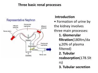
Urinary excretion = Glomerular filtration - Tubular reabsorption + Tubular secretion WHERE & HOW
Tubular reabsorption and tubular secretion • Reabsorption – return of most of the filtered water and many solutes to the bloodstream • About 99% of filtered water reabsorbed • Proximal convoluted tubule cells make largest contribution • Both active and passive processes • Reabsorbed substances carried by the peritubular capillaries to the venous system. • Tubular reabsorption is highly selective (unlike filtration).
Tubular reabsorption and tubular secretion • Secretion – transfer of material from blood into tubular fluid • Helps control blood pH • Helps eliminate substances from the body • Steps • Simple diffusion from peritubular capillaries to interstitial fluid. • Entry to tubular cell can be active or passive. • Exit from tubular cell to lumen can be active or passive.
Tubular Secretion • Tubular secretion is important for: • Disposing of substances not already in the filtrate • Eliminating undesirable substances such as urea and uric acid • Ridding the body of excess potassium ions • Controlling blood pH by secreting H+
Reabsorption and Secretion • filtered load: The amount of a substance filtered into Bowman's space per unit time is called the • Filtered load = GFR [P]x Px = Concentration of substance in plasma • Excretionrate = V [U]x [U]x = Concentration of substance in urine V= volume of urine • Reabsorption/secretion rate = FL – Excretion rate
Reabsorption Routes • Paracellular reabsorption (between the cells) • Between adjacent tubule cells • Tight junction do not completely seal off interstitial fluid from tubule fluid • Passive • Trans cellular reabsorption (across the cell) • Through an individual cell
Reabsorption • Once the substance has moved pass the tubular epithelium cell into the interstitial space, bulk flow then accounts for its movement back into the peritubular capillaries.
Reabsorption - Transport Mechanisms • Transport mechanisms Reabsorption of Na+ especially important • Primary active transport • Sodium-potassium pumps in basolateral membrane only • Secondary active transport • co-transport (glucose, amino acids) • counter-transport (K+, H+) • Passive Reabsorption • Osmosis (H2O) • Electrostatic attraction (Cl-)
Sodium Reabsorption • Na+-K+ ATPaselocated at the basolateral membrane of tubular cells • Creating concentration gradient for Na+ to diffuse into the tubular cells from tubular lumen (diffusion). • Keeps interstitial[Na+] high creating concentration gradient for Na+ to diffuse into blood (Bulk flow).
Glucose Reabsorption • Na+-glucose co transporter in luminal membrane – called SGLT 2 • Proteins involves in facilitated diffusion of glucose at peritubilar membrane is GLUT 2
Additional Examples of Secondary Active Transport These substances include some amino acids, lactate, inorganic phosphate (Pi), H+, and Cl-.
Reabsorption Transport Maximum(Tmax) • Like transport systems elsewhere, renal active transport systems have a maximal rate, or transport maximum (Tm), at which they can transport a particular solute. • As the tubular load increases, all active transport systems becomes saturated, which creates a limit to the rate of substances transport. • Therefore, excess of that substance is excreted.
Glucose Transport Maximum Figure 27-4; Guyton and Hall
RENAL THRESHOLD FOR GLUCOSE • The is the plasma level at which the glucose first appears in the urine . • The actual renal threshold is about 200 mg/dL of arterial plasma, which corresponds to a venous level of about 180 mg/dL. • This deviation is called splay. • What causes in splay ? • TmGin all the tubules is not identical and • All the glucose were not removed from each tubule when the amount filtered was below the TmG.
250 200 Transport Maximum (150 mg/min) 150 Glucose (mg/min) 100 50 0 Filtered Load of Glucose (mg/min) A uninephrectomized patient with uncontrolled diabetes has a GFR of 90 ml/min, a plasma glucose of 200 mg/dl (2mg/ml), and a transport max (Tm) shown in the figure. What is the glucose excretion for this patient? a. 0 mg/min b. 30 mg/min c. 60 mg/min d. 90 mg/min e. 120 mg/min Reabsorbed Excreted . Threshold 50 100 150 200 250 300 350
250 200 Transport Maximum (150 mg/min) 150 Glucose (mg/min) 100 50 0 Filtered Load of Glucose (mg/min) Answer: Filt Glu= Reabs Glu = Excret Glu = (GFR x PGlu) = (90 x 2) = 180 mg/min Tmax = 150 mg/min 30 mg/min GFR = 90 ml/min PGlu = 2 mg/ml Tmax= 150 mg/min Reabsorbed Excreted a. 0 mg/min b. 30 mg/min c. 60 mg/min d. 90 mg/min e. 120 mg/min . Threshold 50 100 150 200 250 300 350
Passive Reabsorption • Passive reabsorption depends on: • Electrical gradient (electrostatic attraction). • Concentration gradient • Membrane permeability • Time available in the tubule for reabsorption
Passive Reabsorption Secondary water Reabsorption via osmosis Sodium reabsorption makes both intracellular and extracellular fluid hypertonic to the tubular fluid. Water follows with sodium into the peritubular capillaries. Na+ Na+ H2O capillary Tubular lumen Tubular cell
Passive Reabsorption Secondary ion reabsorption via electrostatic attraction Negative ions (Cl-) tend to follow with the positive sodium ions by electrostatic attraction. Na Na+ Cl- capillary Tubular cell Tubular lumen
Urea–Example of Passive Reabsorption Na+ reabsorption H2O reabsorption Increase concentration of urea in tubular fluid Passive reabsorption of urea
Mechanisms by which Water, Chloride, and Urea Reabsorption are Coupled withSodium Reabsorption Figure 27-5; Guyton and Hall
PAH –EXAMPLE OF SECRETION • PAH is an organic acid • Used for measurement of renal plasma flow • Both filtered and secreted • PAH transporters located in peritubular membrane of proximal tubular cells. • There are parallel secretory mechanism for secretion of organic bases like quinine and morphine
References • Human physiology by Lauralee Sherwood, seventh edition • Text book physiology by Guyton &Hall,11th edition • Text book of physiology by Linda .s contanzo,third edition