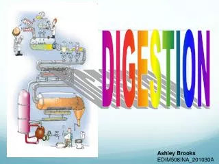
DIGESTION
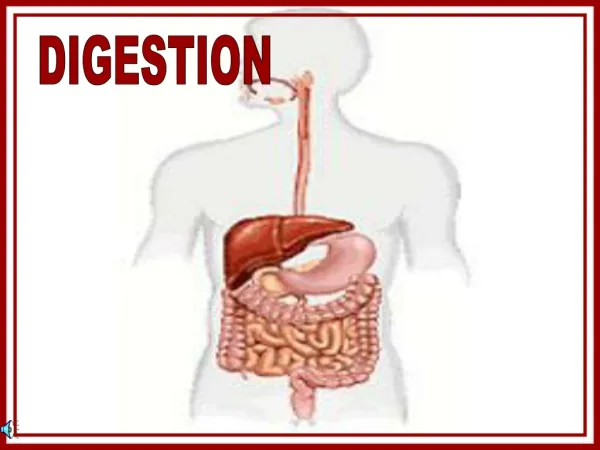
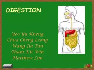
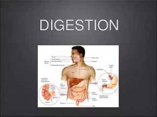
DIGESTION. Digestion. Mechanical and chemical breakdown of foods... Absorption of resulting nutrients by cells ALIMENTARY CANAL: tube extending 9 meters from the mouth to the anus Mucosa—Submucosa—Muscular layer--Serosa LUMEN: space within the intestines. Organs. ALIMENTARY CANAL.

DIGESTION
E N D
Presentation Transcript
Digestion • Mechanical and chemical breakdown of foods... • Absorption of resulting nutrients by cells • ALIMENTARY CANAL: tube extending 9 meters from the mouth to the anus • Mucosa—Submucosa—Muscular layer--Serosa • LUMEN: space within the intestines
Organs ALIMENTARY CANAL ACCESSORY ORGANS Salivary Glands Liver Gallbladder Pancreas • Mouth • Pharynx • Esophagus • Stomach • Small Intestine • Large Intestine • Anal Canal (rectum & anus)
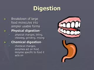
Mouth • Digestion begins here • Mechanical & chemical (starches: amylase) • Lips, teeth, cheek, tongue, salivary glands, papillae, palate, frenulum, tonsils, vestibule, tongue
Tongue • Function: Mix food particles with saliva during chewing and move food toward the pharynx during swallowing • PAPILLAE: bumps on tongue; taste buds • FRENULUM: flap that anchors tongue to bottom of oral cavity
Salivary Glands • Moisten food; secret amylase to begin starch digestion • 3 pairs of major salivary glands • PAROTID glands - largest of the major glands - secretes a clear, watery fluid rich in amylase • SUBMANDIBULAR glands - predominantly serous secretion w/ few mucous cells • SUBLINGUAL glands - smallest of the major glands - secretion primarily mucous type
Teeth • Function: Break pieces of food into smaller pieces • BOLUS: moist ball of food • INCISORS: (8) chisel-shaped with sharp edges to bite off larges pieces of food • CUSPIDS: (4) “canine” teeth; sharp • BICUSPIDS: (8) tear & grind • MOLARS: (12) flattened surface to grind food particles • <Wisdom Teeth> 3rd set of molars; late teens; early 20’s
Parts of Teeth • CROWN: projects beyond the gum • ROOT: anchored to the alveolar process of the jaw • ENAMEL: covers the crown • DENTIN: bulk of the tooth below enamel • PULP: combination of blood vessels, nerves, and connective tissue (blood vessels and nerves reach pulp cavity through ROOT CANAL) • GINGIVA: gum
Pharynx • Connects the nasal & oral cavities with the larynx & esophagus • “back of throat” • 3 parts • nasopharynx: communicates with the nasal cavity & provides a passageway for air during breathing • oropharynx: passageway for food moving downward from the mouth and for air • laryngopharynx: passageway to the esophagus
TONSILS • Produce antibodies to fight infection TYPES LINGUAL • PALATINE • PHARYNGEAL
Uvula • Cone-shaped projection • Function: drawn upward during swallowing to close the opening between the nasal cavity & the pharynx
Esophagus • passageway from the pharynx to the stomach • “food tube” • 25 cm long • PERISTALSIS: muscular contractions that move food • No digestion occurs here • EPIGLOTTIS: flap that closes trachea when we swallow to prevent food/liquid from entering the trachea • LOWER ESOPHAGEAL SPHINCTER: prevents food from backing up into esophagus
Stomach • J-shaped pouch • Just below diaphragm • 1 L capacity or more! • RUGAE: folds • 4 regions: cardiac, fundic, body, and pyloric • CHYME: semifluid paste of food • Chemical digestion of proteins • Gastric juice: HCl & pepsin…highly acidic (pH 2) • PYLORIC SPHINCTER - valve that controls food backing up in the stomach
FUNCTIONS OF THE STOMACH • Begins mixing process with gastric juice • Begins protein digestion • Moves food to small intestine • Limited absorption
Gastric Secretions • Gastric glands contain 3 types of secretory cells: mucous cells, chief cells, & parietal cells = gastric juices • Mucous cells secrete mucus to prevent stomach from digesting itself! • Chief cells secrete digestive enzymes • Parietal cells releases hydrochloric acid • Pepsin: digestive enzyme in gastric juice • Pepsin w/ HCl begins the digestion of nearly all proteins into polypeptide strands • Gastrin: hormone that regulates gastric secretions
Small Intestine • Most important organ of digestion • 6 m if stretched out! • Most absorption takes place here • Many folds (intestinal villi)– increase the surface area for easier absorption • 3 regions: DUODENUM; JEJUNUM; ILEUM
Portions of Small Intestine • Duodenum • C shaped • Receives chyme from stomach • Receives pancreatic juice & bile • Several enzymes released to complete digestion of proteins, dipeptides, disaccharides, fats • Jejunum • Absorption of digested nutrients • Ileum • Absorption of digested nutrients
More about the Small Intestine • MESENTARY: tissue that suspends the jejunum & ileum from the abdominal wall • Lacteal: lymphatic capillary found in the intestinal villi • FUNCTIONS: • Receive secretions from pancreas & liver • Completes digestion • Absorbs products of digestion
Large Intestine • Shaped like an upside down U • 1.5 m long • No villi • Absorbs water & electrolytes • Forms FECES (75% water; undigested material; bacteria; electrolytes • ILEOCECAL SPHINCTER: b/w ileum of small intestine & cecum of large intestine
Regions of Large Intestine • CECUM • ASCENDING COLON • TRANSVERSE COLON • DESCENDING COLON • SIGMOID COLON
Other Info to Know about the Large Intestine • APPENDIX: lymphatic tissue between small & large intestine • HEMORRHOIDS: “pain in the rear”…enlarged/inflammed rectal veins…itching, burning, bleeding • MUCUS produced in colon serves 2 functions: • Binds fecal matter • Protects intestinal wall against abrasive action of undigested waste LARGE INTESTINE VS. SMALL INTESTINE: larger diameter NO VILLI
Rectum & Anus • Feces stored in the rectum • ANAL CANAL: passageway through which feces passes as it passes out the body through the anus • DEFECATION: removal of feces • INTERNAL & EXTERNAL ANAL SPHINCTERS control the release of feces
Liver • Heaviest organ is body (3 pounds) • Well-supplied with blood vessels • Right & left lobes • *blood sugar maintenance • *lipid metabolism (bile secretion) • Produce BILE • *emulsification of fats • *protein metabolism (most important function) • *stores glycogen, iron, vitamins A, B12, D • *removes toxic substances such as alcohol (detoxification)
Stores bile & releases bile to duodenum Released through COMMON BILE DUCT Cholesterol in bile can form crystals (GALLSTONES) Gallbladder
Secretes pancreatic juice Enzymes that digest carbohydrates (amylase), fats (lipases), proteins (trypsin…), & nucleic acids (nucleases) Neutralizes stomach acid Produces insulin Pancreatic duct: connects with duodenum Pancreas