Download
1 / 1
10 likes | 151 Views
The role of fetomaternal MHC class II histoincompatibility in regulating tolerance of the semi-allogenic fetus Darren R. Ritsick, Cassidy Bommer, Jeffrey Braverman Braverman IVF and Reproductive Immunology, PC, NY, USA . Introduction – paternal alloantigen-specific Tregs

E N D
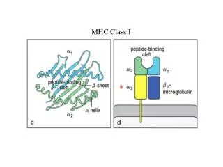
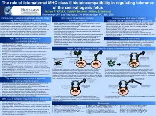
The role of fetomaternal MHC class II histoincompatibility in regulating tolerance of the semi-allogenic fetus Darren R. Ritsick, Cassidy Bommer, Jeffrey Braverman Braverman IVF and Reproductive Immunology, PC, NY, USA Introduction – paternal alloantigen-specific Tregs regulate fetomaternal tolerance MHC class II alloantigens mediate linked suppression Fetomaternal MHC class II disparity regulates linked suppression during pregnancy • Remission from autoimmunity in pregnancy originally thought to owe to effects of hormones and “general” immunosuppression, but now known that fetal antigen-specific mechanisms play an important role in the course of maternal autoimmunity, for better or worse. • Extent of remission of RA, IBD, and MS during pregnancy is correlated with the extent of fetomaternal incompatibility at the HLA class II loci.18,19 • Several autoimmune conditions (gestational dermatosis, systemic sclerosis, thyroiditis) can be triggered during pregnancy by a lack of fetomaternal HLA class II histoincompatibility.20,21 Viviparous pregnancy represents a physiological state in which the maternal immune systemmust tolerate a semi-allogenic fetus expressing ‘nonself’ antigens derived from the paternal genome. A critical role for regulatory T (Treg) cells in mediating active suppression of the maternal immune response to the semi-allogenic fetus is now widely recognized. Significant evidence now exists that that the Treg cells that expand in pregnancy are specific for paternal antigens and function in an antigen-specific fashion. However, roles for specific classes of paternal antigens in the maternal immune response to the fetus are not well defined. • Linked suppression is a mechanism whereby one tolerated antigen is able to confer immune tolerance to a second antigen.14 • The ability to induce linked suppression is a property of MHC class II antigens that is not shared by class I antigens.15 • MHC class II molecules preferentially display MHC class II peptides (pII) that cover polymorphic stretches od allelic sequences derived from hypervariable regions of MHC class II chains.16 • It has been noted that “the propensity of MHC II molecules to present themselves as peptides suggests that pII/MHC II complexes are associated with a specific function”, but that “the cause of preferential presentation of allelic MHC II determinants remains puzzling given that gene allelism has no real meaning within a given mouse strain or human individual” (emphasis added).17 • Indeed gene allelism is rather useful for self/non-self determination amongst individuals of a species. Clinical implications MHC class II molecules regulate reproductive outcome • Lack of significant fetomaternal HLA class II incompatibility may lead to failure to generate tolerance for other classes of paternal antigens by linked suppression. • Therapies that promote linked suppression may be useful to treat recurrent miscarriage in couples significantly lacking HLA class II histoincompatibility. • Paternal alloantigens include MHC antigens (class I and II) and minor H antigens (mHAgs), including H-Y antigens. • MHC diversity is maintained through balancing selection. Pathogen-driven selection is an important mechanism for maintaining MHC diversity, but a role for sexual selection is also accepted. • MHC genes are implicated in both pre- and post-copulatory mate choice in animals.1 • Post-copulatory mate choice (cryptic female choice) mechanisms include selective fertilization, selective implantation, and selective abortion. • Many studies in animals support a role for MHC class II histoincompatibility in reproductive success through both pre- and post-copulatory mechanisms.2 • Early studies on the role of HLA histoincompatibility in humans produced inconsistent results due to highly divergent study methods and low resolution HLA allele typing. • Despite the highly divergent results, a meta-analysis in 2005 concluded that there is a significantly increased risk of spontaneous abortion in couples sharing at least one allele at the HLA-DR locus.3 • Several recent studies using high resolution HLA typing provide further evidence for a significant inverse correlation between HLA class II locus histoincompatibility and the occurrence of recurrent spontaneous abortion and preeclampsia.4-7 Model for role of paternal MHC class II antigens in fetomaternal tolerance Maternal iTreg cell with paternal class II antigen specificity Maternal iTreg cell with specificity for “new” paternal antigen Linked suppression Maternal naïve CD4+ T cells TCR Maternal iTreg cell with paternal class II antigen specificity Maternal naïve T cell with paternal class II antigen specificity Placental exosomes and other placental-derived material containing paternal HLA-DR Paternal class II antigenic peptide Maternal naïve T cell with specificity for “new” paternal antigen not present at conception Soluble and sperm-associated paternal MHC class I, MHC class II, and mHAgs Maternal MHC class II Paternal alloantigen uptake, processing, and presentation Paternal alloantigen uptake, processing, and presentation Maternal tolerogenic APC Paternal antigen newly expressed by fetus during progression of gestation PGE2, TGF-β, and other tolerogenic factors • (1) Paternal class II alloantigens present on sperm and in seminal fluid, in the immune privileged context conferred by factors in the seminal fluid and female reproductive mucosa, are taken up by maternal tolerogenic APCs and presented on MHC class II molecules through the indirect pathway to cause the differentiation of naïve T cells with cognate TCR specificity to iTregs. Although paternal class I and mHAgs are also variably present, initial indirect presentation to maternal CD4+ T cells is dominated by allelic MHC class II peptides by virtue of preferential presentation by MHC class II. • (3) Paternal class II molecules are continually released into maternal circulation by the release of exosomes and other placental-derived material, including apoptotic debris. This maintains tolerance to paternal antigens that were present at the time of copulation, as well as allows for linked suppression to occur for paternal antigens newly expressed by the fetus during progression of gestation (as the class II antigen needs to be co-presented with any “new” paternal antigens by singular APCs). The maternal immune system is exposed to paternal class II antigens Maternal Treg cell with autoantigen specificity Linked suppression • The maternal immune system is initially exposed to paternal antigens through sperm and seminal fluid which contain MHC class II molecules.8 • MHC class II molecules are not expressed at the surface of trophoblasts, but HLA-DR molecules are expressed intracellularly.9 • HLA-DR is sequestered into vesicular compartments, likely by HLA-DO which is expressed by trophoblastsand favors sorting of HLA-DR into exosomes. • Exosomal delivery of antigens to APCs promotes tolerogenicpresentation. • Normal pregnancy is associated with increasing levels of soluble HLA-DR and decreased levels are noted in women with preeclampsia. Maternal naïve T cell with autoantigen specificity Amelioration of maternal autoimmune disease Maternal iTreg cell with paternal class II antigen specificity Maternal iTreg cell with paternal mHAg specificity Linked suppression (2) iTregs with specificity for paternal class II allogenic peptides mediate linked suppression of paternal class I and minor H antigens, thereby inducing a tolerogenic response by the maternal immune system to the full complement of paternal antigens present at that time. • (4) Linked suppression of autoantigens in women with autoimmunity also occurs resulting in amelioration of disease. MHC class II antigens regulate immune tolerance Maternal naïve T cell with paternal mHAgspecificity Maternal iTreg cell with paternal class II antigen specificity • MHC class II histoincompatibility can promote either immune rejection or tolerance depending on context. • MHC class II allele disparity promotes tolerance of tissue allografts transplanted at immunologically privileged sites including the anterior chamber of the eye and the liver.10,11 • Introduction of histoincompatible donor MHC class II molecules into the host in tolerogenic contexts (ie, expression in liver, administration through mucosal routes, and intravenous administration in exosomes) promotes graft tolerance at non-immunologically privileged sites.12,13 References Robertson, S.A., et al., J Reprod Immunol, 2009. 83(1-2): p. 109-16. Ranella, A., et al., Hum Immunol, 2005. 66(1): p. 43-55. Bradley, B.A., et alTransplant Proc, 1995. 27(1): p. 1392-4. Sieders, E., et al., Liver Transpl, 2005. 11(12): p. 1541-9. Cunningham, E.C., et al., Transplantation, 2013. 95(1): p. 70-7. Derbyshire, K., et al., J Immunol, 2011. 186(10): p. 5719-28. Cobbold, S.P. and H. Waldmann, Cold Spring Harb Perspect Med, 2013. 3(6). Ziegler, A., et al., Self Nonself, 2010. 1(3): p. 176-191. Setchell, J.M., et al., Am J Primatol, 2013. 75(10): p. 1021-31 Beydoun, H. and A.F. Saftlas, Tissue Antigens, 2005. 65(2): p. 123-35. Takakuwa, K., et al., Clin Immunol, 2006. 118(1): p. 101-7. Ooki, I., et al., Am J Reprod Immunol, 2008. 60(1): p. 75-84. de Luca Brunori, I., et al., J Reprod Immunol, 2003. 59(2): p. 235-43. Triche, E.W., et al., J Reprod Immunol, 2013. LeGuern, C., et al., J Immunol, 2010. 184(5): p. 2394-400. LeGuern, C., Trends Immunol, 2003. 24(12): p. 633-8. LeGuern, C., Front Biosci, 2007. 12: p. 3133-9. Nelson, J.L., et al., N Engl J Med, 1993. 329(7): p. 466-71. Kane, S., et al., Am J Gastroenterol, 2004. 99(8): p. 1523-6. van Wyk, L., et al., Arthritis Res Ther, 2011. 13(6): p. R183. Chakravarty, E., Arthritis Res Ther, 2012. 14(1): p. 102. Maternal autoantigen