Download
1 / 110
1.11k likes | 1.14k Views
Learn about papulosquamous diseases, including psoriasis, their clinical features, treatment options, and the pathogenesis of these conditions. Explore the epidemiology, genetics, triggering factors, and histology of psoriasis.

E N D
Papulosquamous diseases Dr. Fahad AlSaif Consultant & Associated Professor Chairman of Dermatology Department
Learning Objectives: - Define the papulosquamous disease • Highlight on the pathogenesis of papulosquamous diseases • Discuss the clinical features of papulosquamous diseases • Highlight on the papulosquamous diseases treatment
Papulosquamous disease • The term squamous refers to scaling that represents thick stratum corneum and thus implies an abnormal keratinization process • Papulosquamous diseases are typically characterized by scaly papules
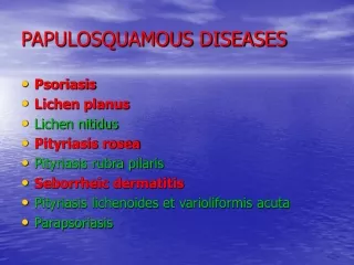
Papulosquamous Diseases: • PSORIASIS • Pityriasis rosea • Lichen planus • Seborrheic dermatitis • Pityriasis rubra pilaris • Secondary syphilis • Miscellaneous mycosis fungoides, discoid lupus erythematosus, ichthyoses
Psoriasis Definition: • is a common, chronic and non-infectious disease. • is a systemic complex disease. Primarily affects skin and joints. • may be a risk factor for metabolic syndrome and its components (abdominal obesity, insulin resistance, hypertension and dyslipidaemia, as well as an independent risk factor for myocardial infarction).
Epidemiology • the disease prevalence remains a questionable : 2% - 4% in adult and 0.5–1% of children • The onset: any age, but two peaks were observed: around 20–30 and over 50 years of age • Pediatric psoriasis: up to 30% of all cases • Race: any race but higher prevalence in western European and Scandinavian populations • low risk in Asians and Africans.
Epidemiology • 75% has nail changes • 30 % of patients with Pso will develop PsA • 75 % of PsA: the skin disease precedes arthritis, while in 15 % of patients Pso appears after PsA and in 10 % the cutaneous and articular involvement are simultaneous
Psoriasis Pathogenesis:- -Who is the pathogenic driver in psoriasis: keratinocyte cells or T lymphocyte cells -Considered to be an autoimmune disease
Psoriasis Pathogenesis -Genetic factor:- -There are two types:-1-type I psoriasis(early onset): more likely to be familial, have a severe clinical course and is associated with HLA-Cw6, B13 and B57 2-type II psoriasis(Late onset ): ages 50 to 60 and is correlated with HLA-Cw2 and B27
several genetic loci for psoriasis have been reported. • There are at least 12 different PSORS loci. • Recently, genome-wide association studies showed 50 regions associated with psoriasis risk.
Psoriasis Pathogenesis -One affected parent: 16%-Both parents :50%-Non-psoriatic parents with affected child: 10%-Monozygotic twins :70% -Dizygotic twins: 20%
Psoriasis Pathogenesis -Environmental factors:-- Infection: streptococcal infection - Physical agents: stress, alcoholism, smoking - Koebner phenomenon - Drugs: lithium, anti- malarials, nsaid, beta-blockers
PsoriasisPathogenesis • Genetically predisposed individuals and triggering factors lead to stressed keratinocytes.(exogenous triggers and indigenous factors) • Stressed keratinocytes will produced:cathelicidin (LL-37) • LL-37 binds to self-DNA and self-RNA released from stressed or dying keratinocytes. • Activates pDCs via TLR9 and TLR7 also activates mDCs via TLR8 • Activated dendritic cells in LN will release IL12, IL23 and TNF-Alfa
Psoriasis Pathogenesis -Epidermal cell kinetics-The growth fraction of basal cells is increased to almost 100% compared with 30% in normal skin -The epidermal turnover time is shortened to less than 10 days compared with 30 to 60 days in normal skin
Psoriasis Histology:-parakeratosis (nuclei retained in the horny layer)-irregular thickening of the epidermis over the rete ridges but thinning over dermal papillae-epidermal polymorphonuclear leucocyte infiltrates (munro abscesses)-dilated capillary loops in the dermal papillae-T-lymph infiltrate in the upper dermis
Psoriasis There are many types of psoriasis:- 1- Non-pustular psoriasis: Psoriasis vulgaris Guttate psoriasis Erythrodermic psoriasis Palmoplantar psoriasis Psoriatic arthritis (PsA) inverse psoriasis 2-Pustular psoriasis Generalized pustular psoriasis (von Zumbusch type) Impetigo herpetiformis Localized pustular psoriasis (Palmoplantarpustular psoriasis and Acrodermatitis continua of Hallopeau
Psoriasis Types 1-plaque psoriasis(psoriasis vulgaris) :- - the most common type. - round-to-oval red plaques and distributed over extensor body surfaces and the scalp - up to 10-20% of patients with plaque psoriasis may evolve into more severe disease, such as pustular or erythrodermic psoriasis
Psoriasis Types 2-Psoriasis, Guttate:- - Small, droplike, 1-10 mm in diameter, salmon-pink papules, usually with a fine scale - Younger than 30 years - Upper respiratory infection secondary to group A beta hemolytic streptococci - On the trunk and the proximal extremities - Resolution within few months
Psoriasis Types 3-ERYTHRODERMIC PSORIASIS:- - Scaly erythematous lesions, involving 90% or more of the cutaneous surface - hair may shed; nails may become ridged and thickened - Few typical psoriatic plaques - Unwell, fever, leucocytosis - excessive of body heat and hypothermia - increase cut blood flow - Increase per-cut loss of water, protein and iron - Increase per-cut permeability
Psoriasis Types • 4-Psoriasis, Pustular:- • - uncommon form of psoriasis • - pustules on an erythematous background • - psoriasis vulgaris may be present before, during, or after • - pustular psoriasis may be classified into several types: • 1-generalized type(von Zumbusch variant): • - generalized erythema studded with interfolecular pustules • - fever, tachypneic, tachycardic • - absolute lymphopenia with polymorph nuclear leukocytosis up to 40,000/µL • 2-Localized form (palms and soles)
Psoriasis Types Causes of pustularps:- Withdrawal of systemic steroids Drugs, including salicylates, lithium, phenylbutazone,, hydroxychloroquine, interferon Strong, irritating topicals, including tar, anthralin, steroids under occlusion, and zinc pyrithione in shampoo Infections Sunlight or phototherapy Cholestatic jaundice Hypocalcemia Idiopathic in many patients
Psoriasis Types 5-Psoriasis inversus(sebopsoriasis):- - Over body folds - The erythema and scales are very similar to that seen in seborrhoeic dermatitis