Download
1 / 55
590 likes | 872 Views
Cell Surface Structures of Bacteria. Other reference. In a prokaryotic cell, the DNA is concentrated in a region called the nucleoid , which is not bounded by a membrane. Procaryotic Form and Function: External Structure.

E N D
Other reference In a prokaryotic cell, the DNA is concentrated in a region called the nucleoid, which is not bounded by a membrane.
Procaryotic Form and Function:External Structure • The evolutionary history of procaryotic cells extends back at least 3.5 billion years. It is now generally thought that the very first cells to appear on the earth were a type of archaea possibly related to modern forms that live on sulfur compounds in geothermal ocean vents. The fact that these organisms have endured for so long in such a variety of habitats indicates a cellular structure and function that are amazingly versatile and adaptable.
Procaryotic Form and Function:External Structure • The general cellular organization of a procaryotic cell can be represented with the following flowchart:
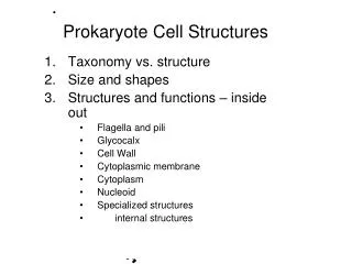
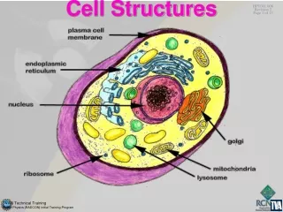
The Structure of a Generalized Procaryotic Cell All bacterial cells invariably have a • cell membrane • ribosomes • cytoplasm • a chromosome • the majority have a cell wall and some form of surface coating or glycocalyx. • Specific structures that are found in some, but not all, bacteria are flagella, pili, fimbriae, capsules, slime layers, and granules (stored nutrients).
The Cell Envelope: TheOuter Wrapping OF Bacteria • The majority of bacteria have a chemically complex external covering (outside of the cytoplasm), termed the cell envelope. It is composed of three basic layers: - glycocalyx - cell wall - cell membrane
The layers of the envelope are stacked one upon another and are often tightly bonded together like the outer husk and casings of a coconut. • Although each envelope layer performs a distinct function, together they act as a single protective unit. • The envelope is extensive and can account for one-tenth to one-half of a cell’s volume.
The Bacterial Surface Coating, or Glycocalyx • The glycocalyx develops as a coating of macromolecules to: - protect the cell from environmental conditions. - in some cases, help it adhere to its environment. • Glycocalyces differ among bacteria in thickness, organization, and chemical composition.: A- Some bacteria are covered with a loose, soluble shield called a slime layer that evidently protects them from loss of water and nutrients.
B- Other bacteria produce capsules of repeating polysaccharide units, of protein, or of both. A capsule is bound more tightly to the cell than a slime layer is, and it has a thicker, gummy consistency that gives a prominently sticky (mucoid) character to the colonies of most encapsulated bacteria.
Specialized Functions of the Glycocalyx • Capsules are formed by a few pathogenic bacteria, such as: -Streptococcus pneumoniae (a cause of pneumonia) - Haemophilus influenzae (one cause of meningitis) -Bacillus anthracis (the cause of anthrax) • Encapsulated bacterial cells generally have greater pathogenicity because capsules protect the bacteria against phagocytes: A capsular coating makes the bacteria too slippery for the phagocyte to capture or digest. By escaping phagocytosis, the bacteria are free to multiply and infect body tissues. • Encapsulated bacteria that mutate to nonencapsulated forms usually lose their pathogenicity.
Other types of glycocalyces can be important in formation of biofilms. The thick, white plaque that forms on teeth comes in part from the surface slimes produced by certain streptococci in the oral cavity. This slime initially allows them to adhere to the teeth and provides a niche for other oral bacteria that, in time, can lead to dental disease. • The glycocalyx of some bacteria is so highly adherent that it is responsible for persistent colonization of nonliving materials such as plastic catheters, intrauterine devices, and metal pacemakers that are in common medical use.
STRUCTURE OF THE CELL WALL • Immediately below the glycocalyx lies the cell wall. • This structure accounts for a number of important bacterial characteristics: - in general, it determines the shape of a bacterium. - it also provides strong structural support necessary to keep a bacterium from bursting or collapsing because of changes in osmotic pressure. (In this way, the cell wall functions like a bicycle tire that maintains the necessary shape and prevents the more delicate inner tube from bursting when it is expanded).
The cell walls of most bacteria gain their relatively rigid quality from a unique macromolecule called peptidoglycan (PG). • This compound is composed of a repeating framework of long glycanchains cross-linked by short peptide fragments to provide a strong but flexible support framework. • Peptidoglycan is only one of several materials found in cell walls, and its amount and exact composition vary among the major bacterial groups.
Other reference The Peptidoglycan Layer • Peptidoglycan is a complex polymer consisting of three parts: 1- a backbone, composed of alternating N-acetylglucosamine and N-acetylmuramic acid 2- a set of identical tetrapeptide side chains attached to N-acetylmuramic acid; 3- a set of identical peptide cross-bridges • The backbone is the same in all bacterial species • The tetrapeptide side chains and the peptide cross-bridges vary from species to species. In many gram-negative cell walls, the cross-bridge consists of a direct peptide linkage between the diaminopimelic acid (DAP) of one side chain and the terminal D-alanine of a second side chain. But in many gram-positive bacteria peptidecross-bridge consists a short glycine amino acids chain. Gram-negative Gram-positive
Other reference • The tetrapeptide side chains of all species have certain important features in common: - Most have L-alanine at position 1 (attached to N-acetylmuramic acid) - Most have D-glutamate or D-isoglutamine at position 2 - Most have D-alanine at position 4. - Position 3 is the most variable one: --- Most gram-negative bacteria have diaminopimelic acid to which is linked the lipoprotein cell wall component. -- Gram-positive bacteriausually have L-lysine; however, some may have diaminopimelic acid or another amino acid at this position. • Diaminopimelic acid is a unique element of bacterial cell walls. It is never found in the cell walls of Archaea or eukaryotes. Diaminopimelic acid is the immediate precursor of lysine in the bacterial biosynthesis of that amino acid
Because many bacteria live in aqueous habitats with a low solute concentration, they are constantly absorbing excess water by osmosis. Were it not for the strength and relative rigidity of the peptidoglycan in the cell wall, they would rupture from internal pressure. • Understanding this function of the cell wall has been a tremendous boon to the drug industry. Several types of drugs used to treat infection (penicillin, cephalosporins) are effective because they target the peptide cross-links in the peptidoglycan, thereby disrupting its integrity. With their cell walls incomplete or missing, such cells have very little protection from lysis. • Some disinfectants(alcohol, detergents) also kill bacterial cells by damaging the cell wall. • Lysozyme, an enzyme contained in tears and saliva, provides a natural defense against certain bacteria by hydrolyzing the bonds in the glycan chains and causing the wall to break down.
Differences in Cell Wall Structure • More than a hundred years ago, long before the detailed anatomy of bacteria was known, a Danish physician named Hans Christian Gram developed a staining technique, the Gram stain, that delineates two generally different groups of bacteria. We now know that the contrasting staining reactions that occur with this stain are due entirely to some very fundamental differences in the structure of bacterial cell walls. The two major groups shown by this technique are the gram-positive bacteria and the gram-negative bacteria. • Because the Gram stain does not actually reveal the nature of these physical differences, we must turn to the electron microscope and to biochemical analysis.
The microscopic section of cell envelopes: • In gram-positive cells, there are two layers: - thick outer cell wall (composed primarily of peptidoglycan) - cell membrane. • In gram-negative cells, there are three layers: -cell wall composed of:- outer membrane - thin layer of peptidoglycan - cell membrane.
The Gram-Positive Cell Wall • The bulk of the gram-positive cell wall is a thick, homogeneous sheath of peptidoglycan ranging from 20 to 80 nm in thickness. It also contains tightly bound acidic polysaccharides, including teichoic acid and lipoteichoic acid. • Teichoic acid is a polymer of ribitol or glycerol and phosphate embedded in the peptidoglycan sheath. Lipoteichoic acid is similar in structure but is attached to the lipids in the plasma membrane. These molecules appear to function in cell wall maintenance and enlargement during cell division, and they also contribute to the H+ ions (acidic charge) on the cell surface directly by H+ release and attraction of H+ by it’s negative group. High [H+] prevent autolysis by enzymes. • In some cases, the cell wall of gram-positive bacteria is pressed tightly against the cell membrane with very little space between them, but in other cells, a thin periplasmic spaceis evident between the cell membrane and cell wall.
The Gram-Negative Cell Wall • The gram-negative cell wall is more complex in morphology because: - it contains an outer membrane (OM), - has a thinner shell of peptidoglycan - has an extensive space surrounding the peptidoglycan. • The outer membrane is somewhat similar in construction to the cell membrane, except that it contains specialized types of polysaccharides and proteins: - The uppermost layer of the OM contains lipopolysaccharide (LPS). The polysaccharide chains extending off the surface function as antigens and receptors. - The innermost layer of the OM is another lipid layer anchored by means of lipoproteins to the peptidoglycan layer below.
Other reference Lipopolysaccharide (LPS) • The LPS of gram-negative cell walls consists of: - a complex glycolipid, called lipid A, -a polysaccharide made up of a core and a terminal series of repeat units (O side chain = O antigen). • The lipid A component is embedded in the outer leaflet of the membrane anchoring the LPS. • LPS, which is extremely toxic to animals, has been called the endotoxin of gram-negative bacteria because it is firmly bound to the cell surface and is released only when the cells are lysed. • When LPS is split into lipid A and polysaccharide, all of the toxicity is associated with the lipid A. The O antigen is highlyimmunogenic in a vertebrate animal. • Antigenic specificity is conferred by the O antigen as this antigen is highly variable among species and even in strains within a species.
The outer membrane serves as a partial chemical sieve by allowing only relatively small molecules to penetrate. • Access is provided by special membrane channelsformed byporin proteinsthat completely span the outer membrane. • The size of these porins can be altered so as to block the entrance of harmful chemicals, making them one defense of gram-negative bacteria against certain antibiotics.
The bottom layer of the gram-negative wall is a single, thin (1–3 nm) sheet of peptidoglycan. Although it acts as a somewhat rigid protective structure, its thinness gives gram-negative bacteria a relatively greater flexibility and sensitivity to lysis. • There is a well-developed periplasmic space surrounding the peptidoglycan.
Other reference The Periplasmic Space • It is space between the inner and outer membranes in gram-negative bacteria. • It contains the peptidoglycan layer and a gel-like solution of proteins. • The periplasmic space is approximately 20–40% of the cell volume. • The periplasmic proteins include: - binding proteins for specific substrates (eg, amino acids, sugars, vitamins, and ions). - hydrolytic enzymesthat break down nontransportable substrates into transportable ones (eg, alkaline phosphatase and 5'-nucleotidase). - detoxifying enzymes that inactivate certain antibiotics (eg, B-lactamase and aminoglycoside-phosphorylase).
Practical Considerations of Differences in Cell Wall Structure Variations in cell wall anatomy contribute to several other differences between the two cell types: • The outer membrane contributes an extra barrier in gram-negative bacteria that makes them more impervious to some antimicrobic chemicals such as dyes and disinfectants, so they are generally more difficult to inhibit or kill than are gram-positive bacteria. One exception is for alcohol-based compounds, which can dissolve the lipids in the outer membrane and disturb its integrity. • Treating infections caused by gram-negative bacteria often requires different drugs from gram-positive infections, especially drugs that can cross the outer membrane.
The cell wall or its parts can interact with human tissues and contribute to disease: - The lipids have been referred to as endotoxins because they stimulate fever and shock reactions in gram-negative infections such as meningitis and typhoid fever. - Proteins attached to the outer portion of the cell wall of several gram-positive species also have toxic properties. For example in: -Corynebacterium diphtheriae (the agent of diphtheria) -Streptococcus pyogenes (the cause of infection of throat) - The lipids in the cell walls of certain Mycobacteriumspecies are harmful to human cells. (Because most macromolecules in the cell walls are foreign to humans, they stimulate antibody production by the immune system)
Nontypical Cell Walls • Several bacterial groups lack the cell wall structure of gram-positive or gram-negative bacteria, and some bacteria have no cell wall at all. • Although these exceptional forms can stain positive or negative in the Gram stain, examination of their fine structure and chemistry shows that they do not really fit the descriptions for typical gram-negative or -positive cells. • For example, the cells of Mycobacterium and Nocardia(an agent of lung or skin infections)contain peptidoglycan and stain gram positive, but the bulk of their cell wall is composed of unique types of lipids. One of these is a very-long-chain fatty acid called mycolic acid, or cord factor, that contributes to the pathogenicity of this group. The thick, waxy nature of these lipids is also responsible for a high degree of resistance to certain chemicals and dyes. Such resistance is the basis for the acid-fast stain used to diagnose tuberculosis and leprosy (caused by Mycobacterium leprae) . In this stain, hot carbol fuchsin dye becomes tenaciously attached (is held fast) to these cells so that an acid-alcohol solution will not remove the dye.
Nontypical Cell Walls - Archaea • Because they are from a more ancient and primitive line of procaryotes, the archaea exhibit unusual and chemically distinct cell walls lacking the true peptidoglycan structure described previously: - In some, the walls are composed almost entirely of polysaccharides. -some have a simple S-layer (Crystalline Surface Layers) comprised of glycoproteins or pure protein. - few archaea lack a cell wall entirely. - some Archaea have a rigid cell wall composed of a peptidoglycan called pseudomurein. Write
The pseudomurein differs from the peptidoglycan (murein) of bacteria: -by having L-amino acids rather than D-amino acids -disaccharide units with an -1,3 rather than a -1,4 linkage. • Archaea that have a pseudomurein cell wall are gram positive.
Some bacteria that ordinarily have a cell wall can lose it during part of their life cycle. These wall-deficient forms are referred to as L forms or L-phase variants (for the Lister Institute, where they were discovered). • L forms arise naturally from a mutation in the wall-forming genes, or they can be induced artificially by treatment with a chemical such as lysozyme or penicillin that disrupts the cell wall. • When a gram-positive cell is exposed to either of these two chemicals, it will lose the cell wall completely and become aprotoplast (a fragile cell bounded only by a membrane that is highly subject to lysis. • A gram-negative cell exposed to these same substances loses it peptidoglycan but retains its outer membrane, leaving a less fragile but nevertheless weakened spheroplast. Evidence points to a role for L forms in certain infections.
CELL MEMBRANE STRUCTURE • Beneath the cell wall • Very thin (5–10 nm) • Bacterial cell membranes contains: - primarily phospholipids (making up about 30–40% of the membrane mass) - - proteins (contributing 60–70%). Proteins account for considerably higher proportion than that of mammalian cell membranes. • The membranes of prokaryotes are distinguished from those of eukaryotic cells by the absence of sterols, the only exception being mycoplasmas that incorporate sterols, such as cholesterol, into their membranes when growing in sterol-containing media (sterols is rigid lipids that stabilize and reinforce the membrane). • Another exception is the membranes of archaea, which contain unique branched hydrocarbons rather than fatty acids. Write
In some locations, the cell membrane forms internal pouches in the cytoplasm called mesosomes. These are prominent in gram-positive bacteria but are harder to see in gram-negative bacteria because of their relatively small size. Mesosomes presumably increase the internal surface area available for membrane activities. Some of the proposed functions of mesosomes are to participate in cell wall synthesis and to guide the duplicated bacterial chromosomes into the two daughter cells during cell division.
Other reference The cell membranes of the Archaea differ from those of the Bacteria: • Some Archaeal cell membranes contain unique lipids, isoprenoids, rather than fatty acids, linked to glycerol by an ether rather than an ester linkage. • Some of these lipids have no phosphate groups, and therefore, they are not phospholipids. Top: an archaeal phospholipid Membrane structures ether linkage phosphate s isoprenoids glycerol phosphate fatty acid Bottom: a bacterial and eukaryotic phospholipid ester linkage
Other reference Write • In other species the cell membrane is made up of a lipid monolayer consisting of long lipids (about twice as long as a phospholipid) with glycerol ethers at both ends (diglycerol tetraethers). The molecules orient themselves with the polar glycerol groups on the surfaces and the nonpolar hydrocarbon chain in the interior. 10 lipid monolayer of some archaea Bottom: 9 lipid bilayer of bacteria and eukaryotes
These unusual lipids contribute to the ability of many Archaea to grow under harsh environmental conditions such as high salt, low pH, or very high temperature.
Photosynthetic procaryotes such as cyanobacteria contain dense stacks of internal membranes that carry the photosynthetic pigments. Functions of the Cell Membrane • Since bacteria have none of the eucaryotic organelles such as mitochondria, the cell membrane provides a site for functions such as energy reactions, nutrient processing, and synthesis. Most enzymes of respiration and ATP synthesis reside in the cell membrane. Enzyme structures located in the cell membrane also help synthesize structural macromolecules to be incorporated into the cell envelope and appendages. Other products (enzymes and toxins) are secreted by the membrane into the extracellular environment.
The glycocalyx and cell wall can bar the passage of large molecules, but they are not the primary transport apparatus. • A major action of the cell membrane is to regulate transport, that is, the passage of nutrients into the cell and the discharge of wastes. Although water and small uncharged molecules can diffuse across the membrane unaided, the membrane is a selectively permeable structure with special carrier mechanisms for passage of most molecules. • The cell membrane is also involved in secretion, or the discharge of a metabolic product into the extracellular environment.
APPENDAGES: CELL EXTENSIONS • Several discrete types of accessory structures sprout from the surface of bacteria. These elongate appendagesare common but are not present on all species. • Appendages can be divided into two major groups: - those that provide motility (flagella and axial filaments) - those that provide attachments (fimbriae and pili).
Flagella—Bacterial Propellers • The primary function of flagella is to confer motility,or self-propulsion. • The extreme thinness of a bacterial flagellum necessitates high magnification to reveal its special architecture, which occurs in three distinct parts: -the filament -the hook (sheath) -the basal body • The filament, a helical structure composed of proteins, is approximately 20 nm in diameter and varies from 1 to 70 nm in length. It is inserted into a curved, tubular hook. • The hook is anchored to the cell by the basal body, a stack of rings firmly anchored through the cell wall, to the cell membrane.
This arrangement permits the hook with its filament to rotate 360°. As the flagellum rotates in a counterclockwise direction, it causes the cell body to swim in a direct forward path. • (it was once thought that the flagellum undulates back and forth like a whip). • One can generalize that all spirilla, about half of the bacilli, and a small number of cocci are flagellated.
Flagella vary both in number and arrangement according to two general patterns: (1) In a polar arrangement, the flagella are attached at one or both ends of the cell. Three subtypes of this pattern are: monotrichous, with a single flagellum; lophotrichous, with small group of flagella emerging from the same site amphitrichous,with flagella at both poles of the cell. (2) In a peritrichous arrangement, flagella are dispersed randomly over the surface of the cell.
The type of arrangement has some bearing on the swimming speed of the bacterium. The speediest forms are polar, flagellated cells such as • Thiospirillum, (5.2 mm/minute) • Pseudomonas aeruginosa (4.4 mm/minute). • Peritrichous rods such as Escherichia coli tend to swim at arelatively slower pace (1 mm/minute).
Fine Points of Flagellar Function • Flagellated bacteria can perform some rather sophisticated feats. They can detect and move in response to chemical signals—a type of behavior called chemotaxis. • Positive chemotaxis is movement of a cell in the direction of a favorable chemical stimulus (usually a nutrient). • Negative chemotaxis is movement away from a repellent (potentially harmful) compound.
Periplasmic Flagella: Internal Flagella • Corkscrew-shaped bacteria called spirochetesshow an unusual wriggly mode of locomotion caused by two or more long, coiled threads, the periplasmic flagella or axial filaments. • A periplasmic flagellum is a type of internal flagellum that is enclosed in the space between the cell wall and the cell membrane. • The filaments curl closely around the spirochete coils yet are free to contract and impart a twisting or flexing motion to the cell.
Appendages for Attachment and Mating • The structures termed pilus and fimbria both refer to bacterial surface appendages that provide some type of adhesion, but not locomotion. We will use the term fimbriae to refer to the shorter, numerous strands and the term pili to refer to the longer, sparser appendages. • Fimbriae are small, bristlelike fibers sprouting off the surface of many bacterial cells. Their exact composition varies, but most of them contain protein.
Fimbriae have an inherent tendency to stick to each other and to surfaces. • They may be responsible for the mutual clinging of cells that leads to biofilms and other thick aggregates of cells on the surface of liquids and for the microbial colonization of inanimate solids such as rocks and glass. • Some pathogens can colonize and infect host tissues because of a tight adhesion between their fimbriae and epithelial cells: - the gonococcus (agent of gonorrhea) invades the genitourinary tract - Escherichia coli invades the intestine by this means. • Mutant forms of these pathogens that lack fimbriae are unable to cause infections.
A pilus (also called a sex pilus) is an elongate, rigid tubular structure made of a special protein, pilin. • So far, true pili have been found only on gram-negative bacteria, where they are involved primarily in a mating process between cells called conjugation, which involves partial transfer of DNA from one cell to another. A pilus from the donor cell unites with a recipient cell, thereby providing a cytoplasmic connection for making the transfer. • Production of pili is controlled genetically, and conjugation takes place only between compatible gram-negative cells.
Other reference Mycoplasmas and Other Cell-Wall-Deficient Bacteria • Mollicutes class(Mollis cutis = soft skin) includes bacteria without cell wall. One member of this class is the Mycoplasmas. • Although other bacteria require an intact cell wall to prevent the bursting of the cell, the mycoplasmal cell membrane (resistant to lysis) is stabilized by sterols and presence of cytoskeletal elemnts
Cytoskeletal Elements • Since mollicutes cells are bounded by only a plastic cell membrane, their dominating shape is a sphere. However, many mollicutes exhibit a variety of morphological entities, including pear-shaped cells, flask-shaped cells with terminal tip structures, filaments of various lengths, and helical filaments. The ability to maintain such shapes in the absence of a rigid cell wall has long indicated the presence of a cytoskeleton in mollicutes. Figure Sectioned images of polarized mycoplasma cells A. M. pneumoniae. B. M. gallisepticum. C. M. mobile.