Download
1 / 74
740 likes | 782 Views
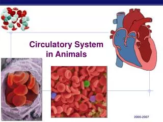
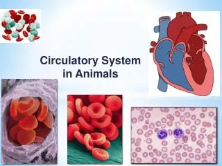
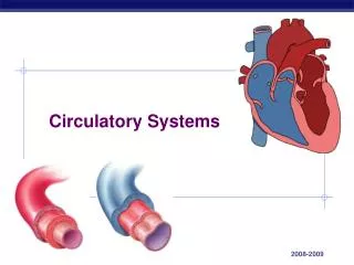
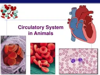
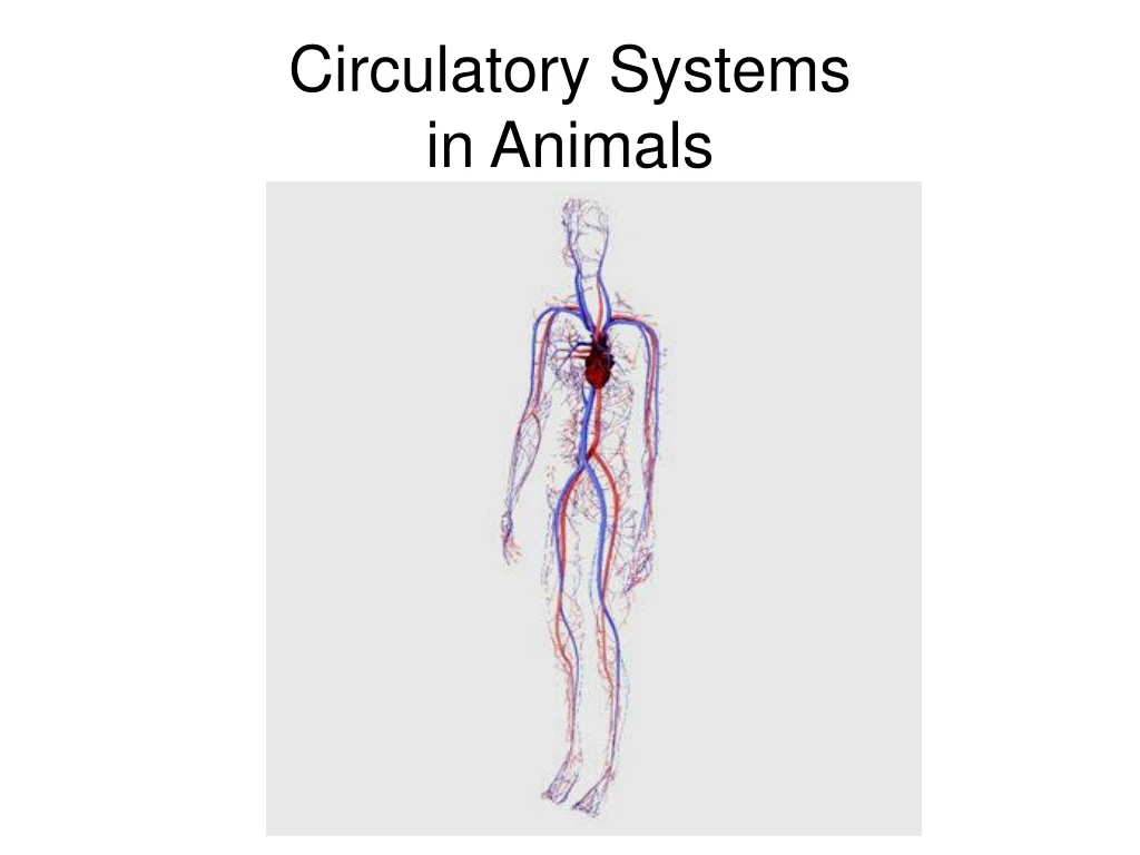
Explore the basics of circulatory systems in animals, including open and closed systems, vertebrate circulatory functions, and the evolution of the vertebrate heart. Learn about the different types of circulatory systems and blood vessels in various animal groups.

E N D
Basic Components of All Circulatory Systems • A fluid, blood, that serves as a medium of transport • A system of channels, or vessels, that conduct the blood throughout the body • A pump, the heart, that keeps the blood circulating
Types of Circulatory Systems • Open Circulatory System example: arthropods and mollusks 2) Closed Circulatory System example: earthworms, vertebrates
Open Circulatory System • Have an open space, the hemocoel • Vessels empty blood into and pick blood up from the hemocoel • Tissues within the hemocoel are bathed directly in the blood
Hemocoel Blood Vessel Blood
Closed Circulatory System • Blood is confined to the heart and a continuous system of blood vessels • Each body cell has a direct blood vessel connection
Functions of the Vertebrate Circulatory System • Transport of oxygen from lungs to tissues • Transport of carbon dioxide from tissues to lungs • Transport of nutrients from digestive system to all body cells • Transport of waste products from all body cells to the liver, then kidney
Functions of the Vertebrate Circulatory System 5. Distribution of hormones from gland to target organ 6. Regulation of body temperature by adjustments in blood flow 7. Prevention of blood loss by means of clotting 8. Protection of body from microbes by circulating white blood cells and antibodies
Evolution of Vertebrate Heart • Two distinctly different chambers: 1) atrium (pl. atria) thin walls and very elastic designed to collect blood from the body 2) ventricle thick walls and very muscular designed to pump blood to the body
Atrium Ventricle
Evolution of Vertebrate Heart • Fish have a 2 chambered heart = one atrium and one ventricle • Amphibians and reptiles have a 3 chambered heart = two atria and one ventricle • Birds and mammals have a 4 chambered heart = two atria and two ventricles
Amphibian, Reptile Fish Bird, Mammal
Fish Circulatory System • Single loop circulatory system • Atrium collects blood from body • Atrium transfers blood to ventricle • Ventricle pumps blood to gills • Gills pick up oxygen and give off carbon dioxide • Blood travels to body cells and gives off oxygen and picks up carbon dioxide
Amphibian and Reptile Circulatory System • Two loop circulatory system • Right atrium collects low oxygen/ high carbon dioxide blood from body • Left atrium collects high oxygen/ low carbon dioxide blood from lungs • Both atria empty into the common ventricle • Deoxygenated and oxygenated blood mix in ventricle and is pumped to body and lungs
Bird and Mammal Circulatory System • Two distinct loops circulatory system • Right loop= pulmonary circulation • Left loop = systemic circulation Deoxygenated blood is always kept separate from oxygenated blood High O2 Low O2 High O2 Low O2
The Blood Vessels • There are 3 types of blood vessels: • Arteries (and smaller arterioles) • Capillaries • Veins (and smaller venules) Vein Artery Capillary
The Blood Vessels: ACV Arterioles Arteries Heart Capillaries Veins Venules
Arteries and Arterioles • Carry blood under pressure away from the heart towards the capillaries • Middle tissue layer of arteries is thick smooth muscle Lumen Thick layer of smooth muscle
Arteries and Arterioles • Smooth muscle contractions regulate blood flow and blood pressure in arteries • Contraction of arteries and arterioles increases blood pressure • Relaxation of arteries and arterioles decreases blood pressure
Capillaries Capillary wall Lumen • Capillaries have cell walls one cell thick easy diffusion of molecules between blood in capillaries and cells in body CO2 O2 RBC Body Cell
Capillary Bed • While individual capillaries are small, capillaries form large capillary beds in tissues very large total area which slows down blood flow increases the rate of diffusion Capillary Bed
Blood Shunting in Capillary Bed • Body regulates the flow of blood through capillary beds • When precapillary sphincters are closed, blood is moved in bulk through a thoroughfare channel called the arteriovenous shunt • This prevents diffusion between blood in capillaries and cells in tissue
Blood Shunting in Capillary Bed • Example: after eating, precapillary sphincters in digestive system capillary beds are open while precapillary sphincters in muscle capillary beds are closed priority is for the blood to pick up the nutrient molecules from digestive system
Veins and Venules • Veins (and smaller venules) drain blood from the capillaries and return it to the heart under low blood pressure • Veins have the same tissue layers as arteries, but have less smooth muscle in the middle layer walls of a vein are thin in comparison to arteries
Connective Tissue Smooth Muscle
Veins and Valves • Theoretically, because veins are thin walled and blood pressure is low in veins, blood should tend to move slowly through the veins • Actually, the flow of blood in the veins increases due to the presence of one-way valves in veins and skeletal muscle contraction around veins
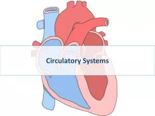
The Human Heart Right Side Left Side • The human heart weighs between 200 to 425 grams and is a little larger than the size of your fist • The heart is located between your lungs in the middle of your chest, behind and slightly to the left of your sternum • The apex of the heart is oriented to the left side of the body
Rat Dissection Left Side Lungs Heart Liver Lungs Right Side Diaphragm
Pericardium • A double-layered membrane sac called the pericardium surrounds the heart • The outer parietal pericardium surrounds the roots of your heart's major blood vessels and is attached by ligaments to your spine, diaphragm, and other parts of your body • The inner visceral pericardium is attached to the heart muscle (myocardium)
Pericardium • A coating of fluid separates the two layers of membrane, letting the heart move as it beats, yet still be attached to your body • This space between the visceral and parietal pericardium is the pericardial cavity
Myocardium • The major portion of the heart is composed of cardiac muscle cells, collectively called the myocardium • Myocardium has a "stringy" look compared to skeletal muscle • Striated skeletal muscle cells are large and lie next to each other in more or less parallel bundles
Myocardium • Cardiac muscle cells are small, butted together at their ends, irregularly shaped, and have numerous blood vessels (BV) between them • Intercalated disks are specialized cell-to-cell adhesion/communications sites and are found only in cardiac muscle.
Heart Chambers • Your heart has 4 chambers • The upper chambers are called the left and right atria ( atrium, singular) and have protruding appendages called auricles • The lower chambers are called the left and right ventricles
Heart Septum • A wall of muscle called the septum separates the left and right atria and the left and right ventricles the right side of the heart is a pump for pulmonary circulation and the left side of the heart is a pump for systemic circulation Left Ventricle Right Ventricle
Heart Valves • The heart has two types of valves: atrioventricular valves and semilunar valves • The tricuspid and mitral (or bicuspid) valves are atrioventricular valves • They have fibrous strands called chordae tendineae on their leaflets that attach to papillary muscles located on the respective ventricular walls • The papillary muscles contract during ventricular contraction and generate tension on the valve leaflets via the chordae tendineae to prevent the AV valves from bulging back into the atria no back flow • The pulmonary and aortic valves are called semilunar valves and do not have chordae tendineae
Blood Flow Through the Heart • The right atrium receives deoxygenated blood from the body via the superior and inferior vena cava • Deoxygenated blood flows from the right atrium, across the atrioventricular tricuspid valve, and into the right ventricle • The right ventricle contracts and pumps deoxygenated blood to the lungs via the pulmonary artery • The semilunar pulmonary valve prevents back flow of the blood into the right ventricle
Blood Flow Through the Heart • Oxygenated blood returns to the heart from the lungs via four pulmonary veins that enter the left atrium • Oxygenated blood flows from the left atrium, across the atrioventricular mitral (or bicuspid) valve, and into the left ventricle • The left ventricle has a very thick muscular wall so that it can generate high pressures during contraction • Oxygenated blood from the left ventricle is pushed across the semilunar aortic valve and into the aorta for transport to the body
Human Circulatory System Two distinct loops circulatory system • Right loop= pulmonary circulation • Left loop = systemic circulation Deoxygenated blood is always kept separate from oxygenated blood High O2 Low O2 High O2 Low O2
Heartbeat and Cardiac Cycle • When surgically removed from the body, the heart will continue to beat for several hours provided it is supplied with the appropriate nutrients and salts • This is possible because the heart possesses its own specialized conduction system and can beat independently even after being separated from its nerve supply • The extrinsic (arising external to the heart) nerve supply coming from the nervous system serves to modify and control the intrinsic (inherent to the heart itself) beating established by the heart
Heart Conduction System Atrioventricular Bundle SA node Inter-nodal Fiber Bundle AV node Purkinje fibers