Download
1 / 59
590 likes | 805 Views
Human Anatomy, First Edition McKinley & O'Loughlin. Chapter 11 Lecture Outline: Axial Muscles. Axial Muscles. Have both their origins and insertions on parts of the axial skeleton. Support and move the head and spinal column.

E N D
Human Anatomy, First EditionMcKinley & O'Loughlin Chapter 11 Lecture Outline: Axial Muscles
Axial Muscles • Have both their origins and insertions on parts of the axial skeleton. • Support and move the head and spinal column. • Function in nonverbal communication by affecting facial features. • Move the lower jaw during chewing. • Assist in food processing and swallowing. • Aid breathing. • Support and protect the abdominal and pelvic organs. • Are not responsible for stabilizing or moving the pectoral or pelvic girdles or their attached limbs.
Muscles of the Head and Neck • Separated into several specific groups. • Almost all originate on either the skull or the hyoid bone.
Muscles of Facial Expression • Originate in the superficial fascia or on the skull bones. • Insert into the superficial fascia of the skin. • Contort the skincausing it to move.
Muscles of Facial Expression • Several are associated with the nose. • The mouth is the most expressive part of the face • muscles in that area are very diverse • Orbicularis oris consists of muscle fibers that encircle the opening of the mouth. • when it contracts the mouth closes
Extrinsic Eye Muscles • Often called extraocular muscles. • Move the eyes. • Are termed extrinsic because they originate within the orbit and insert onto the sclera. • Six extrinsic eye muscles. • the rectus muscles • (medial, lateral, inferior, and superior) • the oblique muscles (inferior and superior)
Muscles of Mastication • Refers to the process of chewing. • Move the mandible at the temporomandibular joint. • Four paired muscles of mastication • temporalis • masseter • lateral pterygoids • medial pterygoids
Muscles That Move the Tongue • The left and right genioglossus muscles originate on the mandible and protract the tongue. • The left and right styloglossus muscles originate on the styloid processes of the temporal bone. • elevate and retract the tongue (pull the tongue back into the mouth) • The left and right hyoglossus muscles originate at the hyoid bone and insert on the sides of the tongue. • Depress and retract the tongue • The left and right palatoglossus muscles originate on the soft palate. • elevate the posterior portion of the tongue
Muscles That Move the Tongue • The tongue is an agile, highly mobile organ. • It consists of intrinsic muscles that curl, squeeze, and fold the tongue during chewing and speaking. • the tongue itself is a big muscle • Extrinsic muscles of the tongue, originate on other head and neck structures and insert on the tongue. • glossus = “tongue” • Used in various combinations to accomplish the precise, complex, and delicate tongue movements required for proper speech. • Manipulate food within the mouth in preparation for swallowing.
Muscles of the Pharynx • Commonly known as the “throat.” • Is a funnel-shaped tube that lies posterior to both the oral and nasal cavities. • Muscles help form or attach to this tube and aid in swallowing. • Primary pharynx muscles are the pharyngeal constrictors (superior, middle, and inferior). • Initiate swallowing and force the bolus inferiorly into the esophagus. • Help elevate or tense the palate when swallowing.
Muscles of the Anterior Neck • The suprahyoid muscles are superior to the hyoid bone. • The infrahyoid muscles are inferior to the hyoid bone.
Anterior and Lateral Neck Muscles • Flex the head and neck downward. • “neck flexion” and “head flexion” refer to the same movement • The main muscles are the sternocleidomastoid and the three scalenes.
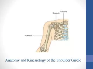
Posterior Neck Muscles • Extend the head/neck. • The trapezius attaches to the skull and helps extend the head/neck. • Primary function is to help move the pectoral girdle.
Muscles of the Vertebral Column • Very complex. • Have multiple origins and insertions. • Exhibit quite a bit of overlap. • Are covered by the most superficial back muscles. • trapezius and latissimus dorsi • The “neck” is the cervical portion of the vertebral column. • The muscles extend the cervical portion of the vertebral column.
Muscles of Respiration • Respiration involves inhalation and exhalation. • During inhalation, several muscles contract to increase the dimensions of the thoracic cavity as the lungs fill with air. • The thoracic cavity expands both to cause the lungs to fill with air and to accommodate the expanding lungs. • During exhalation, some respiratory muscles contract and others relax, collectively decreasing the dimensions of the thoracic cavity and forcing air out of the lungs. • Are on the anterior and posterior surfaces of the thorax. • Are covered by more superficial muscles that move the upper limb.
The Diaphragm • Is an internally placed, dome-shaped muscle. • Forms a partition between the thoracic and abdominal cavities. • The most important muscle associated with breathing. • The muscle fibers converge from its margins toward a fibrous central tendon. • A strong aponeurosis is the insertion tendon for all peripheral muscle fibers.
The Diaphragm • When the diaphragm contracts, the central tendon is pulled inferiorly toward the abdominal cavity, thereby increasing the vertical dimensions of the thoracic cavity. • As it compresses the abdominal cavity, it also increases intra-abdominal pressure. • Also important in helping return venous blood to the heart from the lower half of the body.
Muscles of the Abdominal Wall • Four pairs of muscles collectively compress and hold the abdominal organs in place. • the external oblique • internal oblique • transversus abdominis • rectus abdominis • Work together to flex and stabilize the vertebral column. • When they unilaterally contract they laterally flex the vertebral column.
Muscles of the Pelvic Floor • Formed by three layers of muscles and associated fasciae, collectively known as the pelvic diaphragm. • extends from the ischium and pubis of the ossa coxae across the pelvic outlet to the sacrum and coccyx • Collectively form the pelvic floor and support the pelvic viscera • the pelvic cavity floor is composed of muscle layers that form the urogenital and anal triangles, extend across the pelvic outlet, and support the organs in the pelvic cavity