Download
1 / 33
621 likes | 3.79k Views
Chapter 7: The Biomechanics of the Human Upper Extremity. Basic Biomechanics, 4 th edition Susan J. Hall Presentation Created by TK Koesterer, Ph.D., ATC Humboldt State University. Objectives. Explain how anatomical structure affects movement capabilities on upper extremity articulations.

E N D
Chapter 7:The Biomechanics of the Human Upper Extremity Basic Biomechanics, 4th edition Susan J. Hall Presentation Created by TK Koesterer, Ph.D., ATC Humboldt State University
Objectives • Explain how anatomical structure affects movement capabilities on upper extremity articulations. • Identify factors influencing the relative mobility and stability of upper extremity movements • Identify muscles that are active during specific upper extremity movements • Describe the biomechanical contributions to common injuries of the upper extremity.
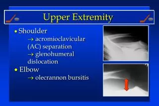
Structure of the Shoulder • Most complex joint in body • Separate articulations: • Sternoclavicular Joint • Acromioclavicular Joint • Coracoclavicular Joint • Glenohumeral Joint • Scapulothoracic Joint • Also: Bursae
Sternoclavicular Joint • Provides major axis of rotation for movement of clavicle and scapula • Freely permitted frontal and transverse plane motion. • Allows some forward and backward sagittal plane rotation. • Rotation
Acromioclavicular Joint • Irregular diarthrodial joint between the acromion process of the scapula and the distal clavicle. • allows limited motions in all three planes. • Rotation occurs during arm elevation • Close-packed position with humerus abducted to 90 degrees
Coracoclavicular Joint • A syndesmosis with coracoid process of scapula • bound to the inferior clavicle by the coracoclavicular ligament. • Permits little movement
Glenohumeral Joint • Most freely moving joint in human body • Glenoid Labrum composed of: • Joint capsule • Tendon of long head of biceps brachii • Glenohumeral ligaments • Rotator Cuff • Rotator Cuff Muscles • Most stable in close-packed position, when the humerus is abducted and laterally rotated.
Scapulothoracic Joint • Region between the anterior scapula and thoracic wall. • Functions of muscles attaching to scapula: • Contract to stabilize shoulder region • Facilitate movements of the upper extremity through appropriate positioning of the glenohumeral joint.
Bursae • Small fibrous sacs that secrete synovial fluid internally to lessen friction between soft tissues around joints. • Shoulder contains: • Subcoracoid bursa • Subscapularis bursa • Subacromial bursa
Movements of the Shoulder Complex • Humerus movement usually involves some movement at all three shoulder joints • Positioning further facilitated by motions of spine • Scapulohumeral Rhythm
Movements of the Shoulder Complex • Muscles of the Scapula • Muscles of the Glenohumeral Joint • Flexion • Extension • Abduction • Adduction • Medial and Lateral Rotation of the Humerus • Horizontal Adduction and Abduction at the Glenohumeral Joint
Muscles of the Scapula • Functions: • 1) stabilize the scapula when shoulder complex is loaded • 2) move and position the scapula to facilitate movement at glenohumeral joint • Are: • Levator scapula, rhomboids, serratus anterior, pectoralis minor, subclavius, and four parts to trapezius.
Muscles of Glenohumeral Joint • Many muscles involved, some contribute more than others. • Large ROM can complicate tension development with orientation of humerus. • Tension development in one shoulder muscle is frequently accompanied by development of tension in an antagonist to prevent dislocation of the humeral head.
Flexion at Glenohumeral Joint • Prime flexors: • Anterior deltoid • Pectoralis major: clavicular portion • Assistant flexors: • Coracobrachialis • Biceps brachii: short head
Extension at Glenohumeral Joint • Gravitational force is primary mover when shoulder extension isn’t resisted. • Control by eccentric contraction of flexors • With resistance there is contraction of muscles posterior to the glenohumeral joint • Assisted by: • Posterior deltoid • Biceps brachii: long head
Abduction at Glenohumeral Joint • Major abductors of humerus: • Supraspinatus • Initiates abduction • Active for first 110 degrees of abduction • Middle deltoid • Active 90-180 degrees of abduction • Superior dislocating component neutralized by infraspinatus, subscapularis, and teres minor
Adduction of Glenohumeral Joint • Primary adductors: • Latissimus dorsi • Teres major • Sternocostal pectoralis • Minor assistance: • Biceps brachii: short head • Triceps brachii: long head • Above 90 degrees- coracobrachialis and subscapularis
Medial and Lateral Rotation of Humerus • Due to action of: • Subscapularis • Has greatest mechanical advantage for medial rotation • Teres major • Assisted by: • Primarily: pectoralis major • Also: anterior deltoid, latissimus dorsi and short head of biceps brachii
Horizontal Adduction and Abduction at the Glenohumeral Joint • Anterior to joint: • Pectoralis major (both heads), anterior deltoid, coracobrachialis • Assisted by short head of biceps brachi • Posterior to joint: • Middle and posterior deltoid, infraspinatus, teres minor • Assisted by teres major, latissimus dorsi
Loads on the Shoulder • Arm segment moment arm: • Perpendicular distance between weight vector and shoulder. • With elbow flexion, upper arm and forearm/hand segments must be analyzed separately. • Large torques from extended moment arms countered by shoulder muscles. • Load reduced by half with maximal elbow flexion
Common Shoulder Injuries • Dislocations • Rotator Cuff Damage • Impingement Theory • Subscapular Neuropathy • Rotational Injuries
Rotational Injuries • Tears of labrum • Mostly in anterior-superior region • Tears of rotator cuff muscles • Primarily of supraspinatus • Tears of biceps brachii tendon • Due to forceful rotational movements • Also: calcification of soft tissues, degenerative changes in articular surfaces, bursitis
Structure of the Elbow • Humeroulnar Joint • Humeroradial Joint • Proximal Radioulnar Joint
Segments at the Elbow • Flexion and Extension • Muscles crossing anterior side of elbow are the flexors: • Brachialis, biceps brachii, brachioradialis • Muscles crossing posterior side of elbow are the extensors: • Triceps, anconeus muscle
Segments at the Elbow • Pronation and Supination • Involves rotation of radius around ulna • Articulations: • Proximal and distal radioulnar joints (both pivot joints) • Middle radioulnar joint (syndesmosis) • Pronator quadratus • Supinator
Loads on the Elbow • Large loads generate by muscles that cross elbow during forceful pitching/throwing • Also in weight lifting, gymnastics • Extensor moment arm shorter flexor moment arm • Tricep attachment to ulna closer to elbow joint center than those of the brachialis on ulna an biceps on radius • Moment arm also varies with position of elbow
Common Injuries to Elbow • Sprains • Dislocations • “nursemaid’s elbow” or “pulled elbow” • Overuse Injuries • Lateral Epicondylitis = “tennis elbow” • Medial Epicondylitis = “Little Leaguer’s Elbow” • Elbow injuries are more chronic than acute
Structure of the Wrist • Radiocarpal joint • Reinforced by: volar radiocarpal, dorsal radiocarpal, radial collateral and ulnar collateral ligaments • Retinacula • Form protective passageways for tendons, nerves and blood vessel to pass through
Movements of the Wrist • Sagittal and frontal plane movements • Rotary motion • Flexion • Extension and Hyperextension • Radial Deviation • Ulnar Deviation
Joint Structure of the Hand • Carpometacarpal (CM) • Metacarpophalangeal (MP) • Interphalangeal (IP)
Movements of the Hand • CM Joints allow large ROM because similar to ball and socket joint • Digits 2-4 constrained by ligaments • MP joints allow flexion, extension, abduction, adduction and circumduction for digits 2-5 • IP joints allow flexion and extension • Extrinsic Muscles • Intrinsic Muscles
Common Injuries of the Wrist and Hand • Sprains and strains fairly common, due to breaking a fall on hyperextended wrist • Certain injuries characteristic of sport type • Metacarpal fractures and football • Ulnar collateral ligament and hockey • Wrist fracture and skate/snowboarding • Wrist in non-dominant hand for golfers • Carpal Tunnel Syndrome
Summary • Shoulder is the most complex joint in the human body. • Movements of the shoulder girdle contribute to optimal positioning of the glenohumeral joint for different humeral movements. • Humeroulnar articulation controls flexion and extension at the elbow • Pronation and supination of forearm occur at proximal and distal radioulnar joints.