Download
1 / 22
220 likes | 401 Views
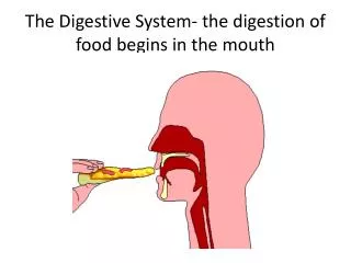
Digestive system of the ox The mouth The cavity of the mouth is shorter and wider-the vestibule is more capacious.

E N D
Digestive system of the ox The mouth The cavity of the mouth is shorter and wider-the vestibule is more capacious. The lips are thick,wide and immobile-middle part of upper lip and the surface between the nostrils is bare-termed muzzle-it is smooth and kept moist by naso-labial glands-form the subcutaneous layer about 1.5cm thick.It shows irregular lines,mapping out small polygonal areas –also a narrow bare strip along the edge of the lower lip-free edge and the adjacent part of the lining membrane bear short,blunt,horny papillae-towards the angles the papillae become longer and sharp pointed.Thelabial glands occur only near the angles forming compact masses. The cheeks are capacious-mucous membrane present large pointed conical papillae directed toward the isthmus faucium – covered with horny epithelium. The buccal glands are very well developed in three parts.Thedorsal part extends from the angle of the mouth to the maxillary tuberosity.Theventral part consist of a compact brownish mass which reaches from the angle of the mouth a short distance under s the massetermuscle.The middle part consist of loosely arranged yellow lobules-small ducts open between the papillae of cheeks. Linear series of large papillae exist on the floor of the mouth on each side of frenumlinguae.Near these are openings of small ducts of sublingual glands.Thecarunculasublingualis,the papilla on which the mandibular duct opens, is wide,hard and has serrated edge.
The hard palate is wide,usuallypigmented.The body of premaxilla is covered with a thick layer of dense connective tissue-thick horny epithelial covering-called the dental plate/pad.The palatine ridges extends backward,about two third of the length of the hard palate-15-19 ridges-ridges are nearly straight and serrated on free edge.A median raphe extends between the ridges.Posterior third of the palate is smooth.Between the dental plate and the first ridge,is the triangular papilla incisiva-on either side of this is a deep furrow,which is the opening of the ductus incisivus-2inches in length and opens on the floor of the nasal cavity-also communicates by a slit like opening with the vomero-nasal organ. The soft palate is shorter but long enough to close the isthmus of faucium.The posterior pillars do not extend to the origin of the oesophagus.Thepalatinus muscle is very much developed.The fibrous aponeurosis is replaced by muscular tissue. The isthmus faucium is wide and dilatable-on either side,behind the anterior pillar of the soft palate,is a deep depression-tonsillar sinus ,lateral to this is the bean shaped tonsil,3-4cm in length.The tonsil does not project into the isthmus faucium,does not occupy the tonsillar sinus and visible internally. THE TONGUE Variably pigmented-root and body are wider-tip is pointed with narrow edge.The posterior part of dorsum forms an elliptical prominence-define infront by a transverse depression-infront there are large and horny filiform and conical papillae with sharp points directed backward-impart rasp-like roughness to the tip and make it efficient in prehension of food.Papillae on prominence are large ,broad and horny-some have blunt,conical forms-others are rounded/flattened-lenticular papillae.
Fungiform papillae are numerous and distinct-scattered over the dorsum and edges of the free part. Valate papillae are about 8-17 on each side-small and form a long,narrow group on either side of the posterior part of prominence of dorsum.Foilate papillae and the lingual fibrous cord is absent. There are lingual follicles in the posterior part of the root and on each side of glosso-epiglottic fold-muscles are well developed-the hyoglossus arises by additional portions from the great and middle cornua of hyoid bone.Tongue is highly protractile and chief organ of prehension. THE TEETH Permanent teeth 2(I 0/4 C0/0 P3/3 M 3/3)=32 Deciduous teeth 2(Di0/4 Dc0/0 Dp 3/3)=20 ERUPTIONS Birth – few days
THE TEETH Permanent teeth 2(I 0/4 C0/0 P3/3 M 3/3)=32 Deciduous teeth 2(Di0/4 Dc0/0 Dp 3/3)=20 ERUPTIONS Deciduous Birth – 2 weeks Di 1/2/3/4 Dp 1 Birth – few days Dp 2/3 Permanent I 1 1.5 – 2 years I 2 2 – 2.5 years I 3 3 years I 4 3.5 – 4 years P 1 2 – 2.5 years P 2 1.5 – 2.5 years P 3 2.5 – 3 years M 1 5 – 6 months M 2 1 – 1.5 years M3 2 – 2.5 years
Eruption of deciduous teeth varies-75% of well bred calves have all incisors erupted at birth. THE SALIVARY GLANDS Parotid gland is smaller and denser-light brown in color-average weight 115gm-form a very narrow long triangular-lies on the posterior part of massetermuscle.The dorsal part is wide and thick-anterior border partly covers the parotid lymph gland.The small ventral end is bent forward-fits into the angle of union of jugular and external maxillary veins-lies on the mandibularglands.The parotid duct leaves the ventral part of the deep face and pierces the cheek-opposite the 5th upper cheek tooth. The mandibular gland is larger than the parotid-pale yellow in color-average weight abot 140gm-covered to a small extent by the parotid-ventral end is large and rounded-related laterally to mandibular lymph gland.The duct leaves the middle of the concave border of the gland-crosses the stylohyoideus and the intermediate tendon of digastricus-ends at the carunculasublingualis. The sublingual gland consist of two parts –dorsal part is long,thin and pale in color-extends from the anterior pillar of soft palate to the symphysis of mandible-has numerous small long tortuous ducts-open between the papillae under the side of tongue.Ventral part is shorter and thicker-lies ventral to anterior portion of dorsal part-pink in color-has a single duct-either opens alongside or joins the mandibular duct.
PHARYNX Short and wide-vault is divided into two cul-de-sac by a median fold of mucous membrane-continuation of the septum nasi-on the lateral wall is small opening of Eustachian tube-covered by simple of mucous membrane.The posterior nares is small.The entrance of oesophagus is large. OESOPHAGUS Shorter,wider and dilatable-average diameter is 5cm-its length is about 90-105cm.Wall is relatively thin-muscular tissue is striped throughout-consist of two strata spiral fibres-except near the stomach-longitudinal and circular-fibres continued into the wall of the stomach for some distance.The mucous membrane forms a prominence on the ventral side of pharyngeal end of tube which contain gland-elsewhere is non glandular. THE ABDOMINAL CAVITY Very capacious-the lumbar part of the spine is about ¼ longer than the horse-the transverse diameter between the last ribs is greater-the costal attachment of the diaphragm rises very steeply from the 8th rib to the dorsal fourth of the last-the abdomen is increased at the expense of the thorax. THE PELVIC CAVITY Narrow and long-inlet is more oblique-elliptical in outline and the transverse diameter is smaller.Pubic part of the floor is horizontal-the ischiatic part slopes dorsally & deeply concave transversely.roof is concave in both directions.The peritoneum extends backward as the 1stcoccygeal vertebra-retroperitoneal part of the cavity is short.
THE STOMACH The stomach is very large-3/4 of the abdominal cavity-fills the left half of the cavity-extends over the median plane into the right half-4 parts-rumen,reticulum,omasum & abomasum-takes its form early in life-division indicated by furrows/constrictions. The first three parts are the proventriculi/oesophagealsacculation-lined with mucous membrane covered by stratified squamous epithelium and non glandular.Theabomasumhas a glandular mucous membrane-true stomach.Theoesophagus open into the stomach – atrium ventriculi.The abomasum joins the small intestine. Large cattle 40-60 gal. Medium cattle 30-40 gal. Small cattle 25-35 gal. Newborn rumen & reticulumhalf as large as the abomasum 10-12 weeks ratio is reverse-omasum is contracted & functionless 4 months rumen & reticulum about 4 times as large as the omasum/abomasum but collapsed and functionless 1.5 years omasum & abomasum similar capacity. Rumen-80%;reticulum-5%;omasum-7/8%;abomasum-7/8%
RUMEN Occupies most of the left half of the abdominal cavity-extends over the median plane ventrally-its long axis reaches from the ventral part of the 7th/8thintercostal space to pelvic inlet-compressed from side to side-2 surface;2 curvatures & 2 extremities. Parietal surface is convex-related to diaphragm,left wall of abdomen and spleen. Visceral surface is irregular-related to the omasum,abomasum,intestines,liver,pancreas,leftkidney,leftadrenal,aorta and the posterior vena cava. Dorsal curvature follows the curve formed by the left part of the crura of the diaphragm and sublumbar muscles-firmly attached by peritoneum and connective tissue as far as the 4th lumbar vertebra. Ventral curvature is convex –lies on the floor of the abdomen-surface are marked by the right and left grooves-indicate externally the division of rumen into dorsal and ventral sacs Reticular/anterior extremity is divided ventrally by a transverse anterior groove into two sacs.The dorsal sac is longer-curves vebtrally over the round,blind end of the ventral sac-is continuous over the reticulum-external line of demarcation- rumino-reticular groove The groove is deep-distinct on part of lateral surface-dorsally no separation exist-rumen and reticulum forming a dome like vestibule which the oesophagus terminates.
Pelvic /posterior extremity extends to pubis-related to intestine and bladder-divided into dorsal and ventral blind sac by a deep transverse posterior groove which connects the longitudinal grooves.The blind sacs are separated from the rumen by the dorsal and ventral coronary grooves. Reticulum is the most anterior and the smallest of the four-is opposite to the ribs from 6th – 7th/8th –the greater part lies on the left f median plane-somewhat piriform but compressed Parietal/diaphragm surface is convex-lies against the diaphragm and liver. Visceral/ruminal surface faces backward-flattened by pressure of the compartments-ends dorsally by joining the wall of the rumen.Thelesser curvature faces to the right and dorsally-connected with the omasum.Thegreater curvature faces to the left and ventrally-lies against the diaphragm-opposite the 6th and 7th ribs. The right extremity/fundusreticuliforms a round cul-de-sac in contact with sternal part of diaphragm,liver,omasum and abomasum-opposite to ventral end of the 6thintercostal space. Omasumis elipsoidal – compressed between the parietal and visceral surfaces-long axis is nearly vertical-lies to the right of median plane-opposite the 7th – 11th ribs inclusive. Parietal surface faces obliquely to the right and forward-related to the diaphragm and liver-below the liver a small area lies against the lateral wall at the ventral part of ribs from 8th-10th ribs-separated by lesser omentum and the diaphragm. Visceral surface faces the opposite direction-in contact with the rumen,reticulum and abomasum.Thegreater curvature faces backward and to the right.Thelesser curvature is short -faces forward and to the left-visible from the left after removal of rumen and reticulum-connected in its upper part with reticulum by a short,narrowneck of the omasum. Below the neck of the omasum- it is crossed by a deep indentations-immediately below is the extensive junction with the abomasum
Abomasumis an elongated sac-lie s on the abdominal floor-anterior blind end, fundus ,is in the xiphoidregion.Thebody of the sac extends backward between the ventral sac of rumen and omasum-turn to the right behind the omasum.Theterminal,smallerpyloric part inclines dorsally-joins the duodenum at the pylorus-usually situated at the ventral part of 9th /10thrib.Theparietal surface is in contact with the abdominal floor-the visceral surface is related to the rumen and omasum.Thegreater curvature gives attachment to superficial part of greater omentum except fundus-adhered to rumen.Thelesser curvature is related to omasum-attached by peritoneum and connective tissue except at the pyloric part-attached to the liver by lesser omentum. The rumen is attached by peritoneum and connective tissue to crura of diaphragm and sublumbar muscles-from hiatus oesophagus backward to 4th /5th lumbar vertebra.A small area of anterior part of visceral surface of rumen is adhered to surface of abomasum.The greater part of lesser curvature of omasum is attached by connective tissue to abomasum. The lesser omentumattaches the ventral part of parietal surface of omasum and the pyloric part of abomasum to visceral surface of the liver. The cavity of the rumen is partially divided into dorsal and ventral sacs pillars-folds of the wall.Theanterior pillar projects obliquely backward and upward from the ventral wall-have a thick concave free edge-opposite to the 11th and 12thribs.The posterior pillar is more horizontal than the anterior pillar-separates the large dorsal and ventral blind sacs.
Its concave anterior border is infront of a transverse plane through the tuber coxae-branches on either side to form the dorsal and ventral coronary pillars –which separate the posterior blind sacs from the general cavity on the sides and ventrally.Theventral coronary pillar is complete-the dorsal coronary pillar fades out.Another branch from the left part of posterior pillar extends forward and upward subsides gradually.Theright pillar have its ventral division fades out about the middle of the surface while the dorsal division joins the posterior pillar.Theleft pillar fades out near the posterior dorsal blind sac.The distance between the middle of anterior and posterior pillars is about 40-45cm in a medium size cow-in this space the dorsal and ventral sacs communicates freely. The anterior end of the dorsal sac of the rumen is separated in its ventral part from the reticulumby a vertical fold-rumino-reticular fold is opposite the 7th /8th rib-free dorsal edge is concave-forms the ventral and lateral margin of the rumino-reticular orifice . The lateral part of the fold fades out lateral to and behind the cardia.The medial part of the fold ends behind the reticular groove and about 7-8cm below the level of cardia-no demarcation of the rumen and reticulum – atrium ventriculi. The cardiais about 10-12cm ventral to the vertebral end of 8th /9th rib-2.5cm to the left of median plane-its position varies by the degree of fullness of rumen and reticulum. The mucous membrane of the rumen is brown-margins of pillars is pale-thickly studded with large papillae about 1cm long.The edges of the pillars and a large part of the wall of the middle of the dorsal sac are not papillated.Thepapillary arrangement is most developed in the blind sacs-vary much in size and form-largest are foliate-many are narrow /filiform-others are conical.The mucous membrane on the medial wall of atrium is wrinkle and non papillated-dorsallyand laterally it is papillated.
Reticular/oesophageal groove begins at the cardia-passes ventrally on the medial wall of atrium and reticulum-end at thereticulo-omasal orifice –about 18-20cm in length-direction is dorso-ventral-the ventral end is about 2.5 – 5cm infront of the plane of the cardia and to the right of the median plane-groove is twisted spirally to the left lip and reverse at the ventral end.The mucous membrane on the lips of the reticular groove is brown and wrinkle-bottom of groove is pale-marked by longitudinal folds-pointed,horny papillae on ventral part. Reticulum –mucous membrane is raised into folds-encloses 4-6 sided cells-term honeycomb –cells are subdivided by smaller folds-bottom are studdedwithpointed,horny papillae-cells grow smaller and gradually disappear near the reticular groove and the edge of the rumino-reticular fold.At the reticulo-omasal orifice– situated in the lesser curvature of the reticulum just to the right of median plane-there are peculiar horny papillae termedunguliform . Omasum – cavity is occupied by hundred longitudinal folds-lamina omasi –spring from the greater curvature and the sides-convex attached edge and a thick,concave free edge which reaches to within a short distance of the lesser curvature.The food is pressed into thin layers in the narrow spaces between the laminae-reduced to fine state by being ground down by numerous rounded,horny papillae which stud the surfaces of the folds.Agroove,sulcusomasi,extends from the reticulo-omasal opening to theomaso-abomasal opening -10cm long-directed ventrally and medially-free from laminae-slight folds and small papillae-function as a direct path from the reticulum to abomasum for fluids and finely divided food.
In the neck of omasum – laminae change to thick folds-a number of peculiar unguliform papillae occur in the lower part of the reticular groove. Theomaso-abomasal orifice is oval-10cm long-bounded infront by thick muscular omasal pillar .The mucous membrane of the abomasumforms an extensive fold on each side of the opening-fold may act as valves-prevent regurgitation of the abomasum. Abomasum – cavity is divided by constriction into two areas-fundus gland region is lined with a soft glandular mucous membrane-which forms extensive spiral folds.Thepyloric region is much narrower.A small cardiac gland zone surrounds the omaso-abomasal orifice .The pyloric orifice is small and round. The lesser omentum leaves the visceral surface of the liver along a line extending from the oesophageal notch to the portal fissures-passes over the parietal surface of the omasum and the pyloric part of the abmasum and the first part of the duodenum. The greater omentumconceals the greater part of the intestines on the right side and covers the ventral sac of rumen-composed of two parts-each composed of two layers of peritoneum-the superficial part extend from the left groove of the rumen ventrally around the ventral sac-ascends on the right side covering the deep part-ends along the second part of the duodenum and the greater curvature of the abomasum-the deep part is attached along the visceral surface of the rumen ventral to the right groove-curves around the intestinal mass to the right side-blends with the medial layer of the mesoduodenum-anteriorly is attached on the first bend of colon and the visceral surface of the liver along the ventral border of pancreas.The two parts are continuous at the attachment along the posterior groove of the rumen-also join at the iliac flexure of the duodenum and at the origin of the colon-the epiploic foramen ia almost sagittal in direction.
Muscular coat of rumen consist of two layers-fibres of the external layer are longitudinal-the thick internal layer are largely circular – scattered bundles of striped muscles radiate from the cardia in wall of atrium and along the reticular groove. Muscular coat of reticulum consist of two layers-begin and end at the oesophageal groove-pass in a circular or oblique around the sac-fibres of the two layers crossing each other at varying angles.The lip of the reticular groove consist of a thick layer of longitudinal fibres-largely continuous above the cardia-the right spread out ventrally as internal fibres of reticulum-the left lip go to the omasum.The bottom of the groove has two layers of oblique,unstriped muscle-fibres with a variable outer layer of striped-muscle continuous with the oesophagus. Muscular coat of omasumconsist of a thin external longitudinal layer and a thick internal circular layer.At the omasal there is an incomplete inner layer of oblique fibres. Muscular coat of abomasumconsist of longitudinal and circular layers-forms the pyloric sphincter.Mucous membrane is covered with a thick,stratified,squamous epithelium-shed in large patches in rumen and omasum-tunica propria is papillated and glandular.The mucosa of the fundus gland is very thin and thicker towards the pylorus Vessels and nerves –blood supply from the coeliac artery and the veins to go to portal vein-nerves from vagus and sympathetic.
The Intestines Caudal of the alimantery canal – commences at the pylorus and continues to the anus. The small intestine from the pylorus to the caecum . The large intestine from the caecum to the anus. Small intestine : a.theduodunum b.the jejunum c.the ileum Large intestine : a.the blind caecum b.the colon c.the rectum Generally the length of the intestine : a.Five times the body length of carnivorous b.ten times the body length in the horse c.Twenty times the body length in ruminants
Structure of the intestine walls a.Mucosa b.Submucosa c.Muscular layer d.Peritoneum The single-epithelium of the mucosa consists of columnar cells-functional in absorption-the surface mucosa increased by the presence of innumerable intestinal villi-densely packed,finger-like projections give the luminal surface of the small intestinal its velvety look. The mucosa of the large intestine have no villi-the intestinal glands of the large intestine are longer,straighter and richer in goblet cells-produce the mucus to ensure a smooth passage-its important function is the re-absorption of water and the hydration of its faecalcontents.The lymphatic tissue of the intestinal wall is the first line of defence against microorganisms – present as scattered lymphoctyes in the mucosa-single follicles or may aggregate to form Peyer patches-Peyer patches of the mucosa as irregularly plaques-In Ox in the ileum and large bowels of the ruminants. The submucosa consists of loose connective tissue with smaller blood vessels,lymphatics and lymph follicles. The muscular layer consists of a thin outer longitudinal layer and a thicker inner circular layer. The serosal layer of the intestine is provided by the visceral part of the peritoneum.
Duodenum Duodenum can be subdivided into: a.Cranial portion b.Cranial duodenum flexure c.Descending portion d.Caudal duodenum flexure e.Ascending portion f.Duodenumjujenal flexure The portion continues from the pylorus of the stomach and passes towards the right abdominal wall before the deflecting caudally to descend to the pelvic inlet-passes medially around the cranial root of the mesentery before the ascending cranially ,end by bending ventrally .The caudal end of the duodenum is marked by the cranial border of the duodenocolicfold.The duodenum is attached to the abdomen root by the mesoduodenum,the cranial part of the mesentery. The cranial part of the duodenum is connected with the liver by the hepatoduodenalligament.Within the hepatoduodenal ligament passes the common bile duct from the liver to the duodenum-mesentery of the descending duodenum includes the right lobe of the pancreas.
Jejunum The longest part of the small intestine-the most mobile due long to the mesojejunumwhihsupends the jejunum and ileum from the abdominal root. The mesojejunum is continuous with the mesoileum –form of the large fan hanging from the abdominal root with the convoluted jejunum and ileum –the very sort,bunched portion at the aorta as known root of the mesentery – cranial mesentery artery,the large mesentery plexus of nerves that surround the artery and intestinal lymphatics. The large rumen occupies the left half of the abdomen-pushing the intestines to the right.The ascending colon is partly to the right surface of the mesojejunum-Lymphoid tissue is generously spread throughout the mucosa. Ileum The short terminal part of the small intestine-the distinction between the jejunum and the ileum is the ileocaecum fold-it ends at the ileocaecum junction with the ileal opening on the ileal papilla. Large intestine Can be subdivided : a.Caecum b.Colon – ascending colon – transverse colon – descending colon c.rectum
Caecum The first of the large intestine-blind tube and communicates with the ileum through the ileal orifice and with the colon through the caecocolic orifice. The ruminant have small caecum and have neither taenia nor haustra-is located in the right half of the abdomen within the superomental recess with its blind apex pointing caudally. Colon Three parts: a.Ascending colon b.Transverse colon c.Descending colon The colon is divided into the usual ascending,tranverse and descending parts-the ascending colon is the far longest and has a characteristic spiral arrangement. After the caecum the ascending colon forms a sigmoid flexure,first cranially convex and the second caudally convex-narrows and turns ventrally to form a double spiral – with the left side of the mesentery-there are four centripedal followed by the same number of the centrifugal turns-the centrifugal turns of small ruminants have a pearl string appearance due to the characteristic of the faeces. After the last centrifugal loop of the spiral the ascending colon continues into a distal loop and away from the pelvic to join the transverse colon.The short transverse colon crosses from the midlife cranial to the mesenteric root and caudally as the descending colon.
Rectum The descending colon enter the pelvis become the rectum-most of the rectum is suspended by the mesorectum but the terminalyretroperitoneum-the retroperitoneum space is filled with soft tissue rich in fat. Gland associated with the alimentary canal Liver The largest gland in the body – both the exocrine and endocrine in function.Bile is stored and concentrated in the gall bladder-bile is responsible for emulsifying the fatty compound prior to absorption.Endocrine substance are release into the blood stream and play an important role in the fat,carbohydrate and protein metabolism. The liver is located in the thoracic part of the abdomen-the bulk of the liver lies to the median plane-the rumen pushed the liver into the right half of the abdomen.It has a strongly convex surface towards the diaphragm and a concave surface facing the towards other abdomens.The visceral surface is marked by the hepatica porta through which the portal vein,bile duct and the hepatic vessels enter/leave the organ. The liver of the ruminants has no fissures-consists of a left and right lobe,a quadrate lobe and caudal lobe.
Bile ducts The bile is produced by the sheets of hepatocytes and discharged intothe bile canaliculi-capillaries unite to form the interlobular ducts-the interlobular ducts to forms the lobar ducts. The extrahepatic bile ducts consists of the hepatic ducts from the liver,the cystic duct to the gall bladder and the bile duct to the duodenum.In the ruminants the labor ducts unite to form a left and right hepatic duct,which again unite to form the common hepatic ducts. Gall bladder The sac-like gall bladder lies in a fossa on the visceral surface of the liver close to the hepatic porta-it stores the bile and discharges it into the duodenum-also concentrates the bile by absorption through the folded mucosa-a gall bladder is not present in the horse. Pancreas Pancreas has both an exocrine and endocrine function.Its exocrine pancreatic juice is conveyed to the dudenum-its three enzymes for the reduction of proteins carbohdrates and fats.The endocrine part of the pancreas produces insulin,glucogen and somatostatin.
The pancreas is located in the dorsal part of the abdominal cavity to the proximal part of the duodenum-divided into three parts : a.body of the pancreas b.right lobe of the pancreas c.left lobe of the pancreas The pancreas of ruminants consists of a short body and right and left lobes-the right lobe is larger and follows the mesentery of the descending part of the duodenum-the portal vein passes over the dorsal border of the organ in the pancreas notch. A pancreas commonly drains the part of the gland that arises from the ventral primordium and opens into the duodenum together with the bile duct on the major duodenal papilla. Pancreas duct sytem of the different species a.Cat :pancreas duct large,small accessory pancreas duct present in some individual b.Dog :pancreas duct small,missing in some individuals,accessory pancreas duct large. c.Pig :accessory pancreas duct d.Ox :pancreas duct extremely rare,accessory pancreas duct e.Small ruminants :pancreas duct,some sheep with accessory pancreas duct f.Horse :pancreas duct large,accessory pancreatic duct small.