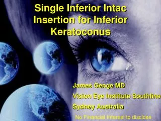
Keratoconus
Keratoconus . Dr. Abdullah S. Al Yousef. Definition. A non-inflammatory eye condition in which the normally round dome-shaped cornea progressively thins causing a cone-like bulge to develop. This results in significant visual impairment. Pathophysiology.


Keratoconus
E N D
Presentation Transcript
Keratoconus Dr. Abdullah S. Al Yousef
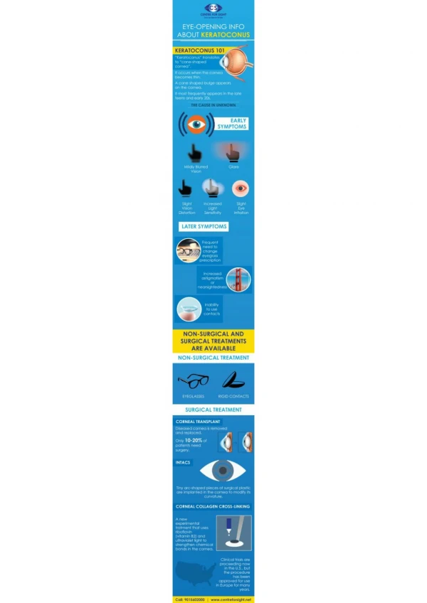
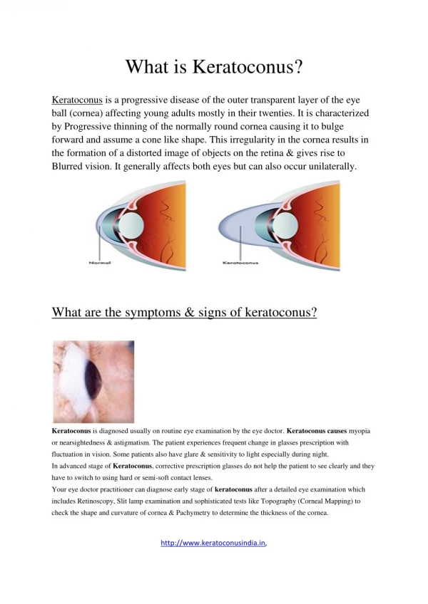
Definition A non-inflammatory eye condition in which the normally round dome-shaped cornea progressively thins causing a cone-like bulge to develop. This results in significant visual impairment.
Pathophysiology • All layers of the cornea are believed to be affected by KC, most notable features are the. ◦1. Thinning of the corneal stroma. ◦2. Ruptures in the Bowman layer. ◦3. Deposition of iron in the basal epithelial cells, forming the Fleischer ring. ◦4. Breaks in and folds close to the Descemet membrane result in acute hydrops and striae, respectively
Symptoms • Blurred Vision • Myopia , Astigmatism • Glare at night • Photophobia • Diplopia
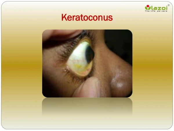
Signs • Corneal thinning • Fleischer's ring (an iron colored ring surrounding the cone) • Vogt's striae (stress lines caused by corneal thinning) • Apical scarring (scarring at the apex of the cone) • Munson's sign: It’s an angulation of the lower lid during inferior gaze due to corneal protrusion • Photokeratoscope with normal round curvature
Measurements of corneal thickness and curvature • The most commonly used approach is ultrasonic pachymetry. the probe must touch the corneal surface and topical anesthesia is thus required. Its accuracy is dependent on the perpendicularity of the probe's application to the cornea and reproducibility relies on precise probe placement on the corneal center. • The Orbscan II corneal topography system (Bausch & Lomb) is an optical scanning-slit instrument that provides topographic analysis and pachymetric measurements of the cornea. • This disparity between instruments can result from their distinct methodologies. The noncontact Orbscan system measures the hydrated mucous component of the tear film over the cornea; contact ultrasonic pachymetry does not. Thus, Orbscan readings are higher than ultrasonic readings.
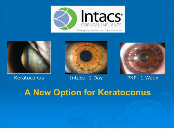
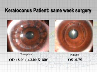
Treatment • To improve vision & to stop progression • Glasses or soft contact lenses in mild or early keratoconus. • Rigid gas permeable lenses in the intermediate stages. • Advancing keratoconus cant tolerate rigid contact lens • New treatments: • Corneal ring -Intacts corneal rings (placing with the corneal stroma in the periphery of the cornea. The result is a flatter cornea and clearer vision.) • C3R - Corneal Collagen Crosslinking with Riboflavin. Increasing collagen crosslinking within the cornea. Combined with Intacs to provide a combined effect and provide greater stability than one treatment alone. • Cornea transplant = penetrating keratoplasty. A donor cornea will replace the thinning cornea and can often provide stable vision. Patient will most likely need glasses or contact lenses for clear vision