Download

1 / 12
120 likes | 161 Views
A 32-year-old man with keratoconus developed corneal melting post-CXL due to Acanthamoeba infection, leading to corneal perforation necessitating therapeutic keratoplasty. This unique case highlights potential risks of CXL and discusses factors contributing to corneal melting. The role of deepithelialization, topical agents, and underlying infections are explored.

E N D
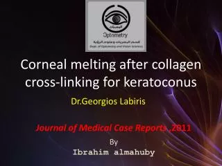
G. PAGANONI – P. RAMA SAN RAFFAELE SCIENTIFIC INSTITUTE MILAN Authors have no financial interest CORNEAL PERFORATION AFTER CROSSLINKING TREATMENT FOR KERATOCONUS
A 32-year-old man with keratoconus developed corneal melting five days after CXL. Corneal scraping was positive for Acanthamoeba. Due to corneal perforation a large therapeutic keratoplasty was performed. Although considered a safe procedure this case emphasizes the potential risks involved in CXL. We discuss the potential effects of deepithelialization, contact lens placement, topical NSAIDs and anesthetics instillation, and the possible role of apoptosis and denervation when performing CXL. ABSTRACT
32-year-old man good general health bilateral keratoconus since 1993 CL discontinued due to intolerance visual acuity worsened in left eye in the last year keratoconus progression confirmed by 2 consecutive topography tests optical pachymetry > 400 μm no other ocular pathology PATIENT
February 2008: CXL (Siena Eye Cross protocol) 9 mm epithelial removal 15 minutes riboflavin (Ricrolin) instillation 30 minutes UVA irradiation (CBM VEGA – CSO) riboflavin instillation every 2' during irradiation Medication with: ofloxacin drops x3 flurbiprofen drops x4 artificial tears BANDAGE CONTACT LENS CXL PROCEDURE
DAY 1 patient noticed redness and conjunctival discharge rinsed his eyelids with TAP WATER to remove secretions referred to ophthalmologist: was added levofloxacin x4 DAY 3 Corneal involvement with opacification and ulceration Therapy prescribed: ofloxacin x5 levofloxacin each hour flurbiprofen x3 chloramphenicol + betamethasone ointment x4 ceftriaxone i.m. 4 g/day prednisone p.o. 25 mg/day DAY 5 Condition worsened Hospitalization and conjunctival flap scheduled
DAY 6 Patient referred to San Raffaele Hospital, presented with: severe inflammation subtotal epithelial defect corneal ectasia corneal infiltrate and opacification ulceration and melting
Corneal scrapings for bacteria, fungi, Acanthamoeba and herpes virus performed Smears were positive for Acanthamoeba Cultures resulted successively negative for all microorganisms Prescribed therapy: acyclovir 800 mg x3/day ceftazidime i.m. 1 gr x2/day levofloxacin every 2 hours hexamidine every 2 hours PHMB every 2 hours
DAY 11 Corneal perforation occurred Therapeutic 10 mm PK performed Histology of the corneal button showed loss of Bowman and Descemet membranes, stromal necrosis and severe granulocytic infiltrate No Acanthamoeba, bacteria and fungi found
Can provoke inflammation, melting and corneal perforation A pre-existing Acanthamoeba keratitis seems unlike: patient did not wear CL before CXL Soft CL contamination with tap water is a well known risk factor Acute infections are possible BUT: Rapid progression of corneal melting is unusual even after PRK Wagoner: “Acanthamoeba keratitis after photorefractive keratectomy” JCRS 2002 ACANTHAMOEBA
Remain uncertain Sings of bacterial infection were referred since post-op day1 BUT: No bacteria, fungi nor herpes found on smears and cultures Only Acanthamoeba was present in smears Histology was negative Possible risk factor are: infections (bacteria – Acanthamoeba) epithelial debridement use of topical NSAIDs corneal toxicity from topical anesthetics denervation keratocyte apoptosis? ETIOLOGY OF CORNEAL MELTING
A presumed bacterial infection, in association with the lack of integrity of the epithelium, denervation and keratocyte apoptosis secondary to UVA irradiation, the frequent instillation of topical anesthetics during the procedure, NSAIDs instillation and Acanthamoeba infection may have led to the activation of multiple noxious mechanism that caused ulceration and corneal melting. SUMMARY
A risk of microbial keratitis exist in CXL due to epithelial defect. The post-operative use of bandage contact lens and NSAIDs (not contempled in the original treatment) should be well evaluated. The role of denervation and keratocyte apoptosis following CXL has still to be investigated. CONCLUSION