Download
1 / 8
90 likes | 255 Views
Idenfitied Differentially Expressed Genes in Keratoconus. Ji Eun Lee, Jong Soo Lee Department of Ophthlamology, Graduate School of Medicine, Pusan National University. Authors have no financial interest in any materials discussed in this article. Purpose.

E N D
Idenfitied Differentially Expressed Genes in Keratoconus Ji Eun Lee, Jong Soo Lee Department of Ophthlamology, Graduate School of Medicine, Pusan National University 20071116_mtDNA workshop
Authors have no financial interest in any materials discussed in this article. 20071116_mtDNA workshop
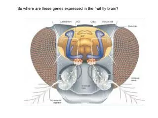
Purpose • It is valuable to examine the genes of the keratocytes that are involved in the thinning of the cornea because investigation of the fundamental pathogenesis is important to understand the disease of the keratoconus. • The purpose of this study is to evaluate the pathogenesis of keratoconus through differentially expressed genes in human keratocyte of keratoconus.
Methods • Culture of human keratocyte • mRNA isolation • cDNA synthesis • Annealing Control Primer (ACP)-based GeneFisingTM PCR • Cloning and Sequencing • RT-PCR • Quantitative Real-Time PCR Figure 1. ACPTM structure. APCTM technology provides a primer with annealing specificity to the temple and allows only real products to be amplifies, such that it enables the researchers to find only real products as a result. A: core sequence - annealing at the first stage of PCR (targeting). B: universal sequence - annealing at the second stage of PCR. C: regulator- regulating the functions of A and B.
1 2 1 2 1 2 1 2 500bp 500bp 1000bp 1000bp A B C D A B A E A 1 2 1 2 1 2 1 2 1 : 정상각막 2 : 원추각막 F G H E Results Figure 2. ACP-based PCR product of the eight verified differentially expressed genes showing increased or decreased levels of expression in keratoconus (arrows). 1: normal cornea. 2: keratoconus. A: BMP4. B: ACTA 2. C: CFL 1, D: GRCC 10, E: TIMP 3, F: MRVI1, G: TIMP1, H: SSTR1.
1 2 1 2 1 2 ACTA2 CFL1 BMP4 GAPDH GAPDH GAPDH 1 2 1 2 GRCC10 TIMP3 GAPDH GAPDH 1 2 1 2 1 2 MRVI1 TIMP1 SSTR1 GAPDH GAPDH GAPDH Results Figure 3. Depicted are RT-PCR products of the eight verified genes and a housekeeping gene (GAPDH). 1: normal cornea. 2: keratoconus. • Cytoskeleton - CFL1, GRCC10, SSTR1, TIMP1, TIMP3 • Wound healing - ACTA2 • Apoptosis- BMP4, CFL1, MRVI1
Results Figure 4.Quantitative real-time polymerase chain reaction. Quantitative real-time polymerase chain reaction of the eight verified genes using GAPDH as endogenous control showed that BMP4 (A), CFL1 (B), MRVI1 (C) were increased by 1.6, 3.3, and 11 fold, respectively, whereas ACTA2 (D), GRCC10 (E), TIMP3 (F), TIMP1 (G), and SSTR1 (H) were decreased by 4.5, 2.7, 14, 8.5, and 1.8 fold, respectively in keratoconus relatively to norma l cornea.
Conclusions • We found differential expression of genes related to apoptosis, as well as those related to cytoskeleton structure and wound healing in keratoconus, using GeneFishingTM PCR. • This was based on the PCR method using the mRNA of keratocytes in normal cornea and keratoconus. • We also confirmed the importance of the apoptosis, cytoskeleton, and wound healing of keratocytes as an important cause of keratoconus through the differential expressions of genes. • We anticipate that gene therapy techniques using such genes will suppress the progress and side effects of keratoconus in the future.