Download
1 / 1
10 likes | 197 Views
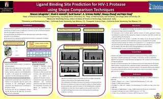
PDB. Largest Pocket Vol/ Ligand Vol. Dipole Moment Len. Inertia. Quarduple moment. p_Integral. Betti Numbers. Protein1B6J. 862.77362. 11.0749. 151.16163 141.32252 27.399763. 123.522423 -22.102247 -101.420181. -2910.009. 1 4 1. 0 0 0. Ligand PI1. 354.51686. 0.00000.

E N D
PDB Largest Pocket Vol/ Ligand Vol Dipole Moment Len Inertia Quarduple moment p_Integral Betti Numbers Protein1B6J 862.77362 11.0749 151.16163 141.32252 27.399763 123.522423 -22.102247 -101.420181 -2910.009 1 4 1 0 0 0 Ligand PI1 354.51686 0.00000 33.238232 30.054001 6.967624 49.356991 -19.902149 -29.454842 354.51690 0 6 0 11011 Ligand Binding Site Prediction for HIV-1 Protease using Shape Comparison Techniques Manasi Jahagirdar1, Vivek K Jalahalli2, Sunil Kumar1, A. Srinivas Reddy3, Xiaoyu Zhang4 and Rajni Garg5 1Dept. of Electrical And Computer Engineering, San Diego State University, CA, 2Dept. of Mathematics and Statistics, San Diego State University, CA 3Molecular Modeling Group, Indian Institute of Chemical Technology, Hyderabad, India 4Chemistry and Biochemistry Dept., California State University, San Marcos, CA, 5Computer Science Dept., California State University, San Marcos, CA Introduction Description Discussion • Effective binding site prediction is a primary step in the molecular recognition mechanism and function of a protein with an application in discovery of new HIV protease inhibitors that are active against mutant viruses • Accuracy of binding-site prediction can be improved using a combination of shape descriptors for the interfaces • We use geometrical, topological and functional descriptors in combination for ligand binding site prediction of HIV-1 protease • The dataset for the algorithm for binding site prediction and extraction : 90 HIV protease protein (21 wild type, and 69 mutated) PDBs • The descriptors such as volume, dipole moment, moment of inertia, quadruple moment, hydrophobicity, residue interface propensity, integral of properties, and, Betti numbers are used for predicting the binding site • The largest pocket of the protein is invariably the binding site for the ligand and hence residue interface propensity and hydrophobicity values are calculated for this pocket • Predicted interface residues are residues with propensity >= 1.5. A propensity of 0 indicates that the amino acid has the same frequency in the interface and surface area • For this dataset, ALA, ASP, ARG and VAL have high preference in the interface • Predicted interface residues are distinctly hydrophobic. Dataset: Mutated and Wild Proteins Residue Interface Propensity Values 3D visualisation of protein, pocket and ligand and descriptor information • PDB : 1B6J • Mutation : C67ABA, C95ABA, C167ABA, C195ABA • 1B6J is a HIV protease complexed with macrocyclic peptidomimetic inhibitor Protein Residue propensity and Hydrophobicity results for protein pocket Pocket Ligand Future Work Binding Site • Research and statistical results has proved the importance of utilizing a combination of descriptors in predicting binding sites of proteins. In the future, we plan to extend the algorithm to include more shape descriptors like tightness of fit, curvature in fine tuning the binding site prediction • We plan to study the alternative sites for binding and the role of the attributes like volume, dipole moment, moment of inertia, quadruple moment, hydrophobicity, residue interface propensity, integral of properties, and, Betti numbers in the alternate binding site prediction • This study can be extended for other HIV targets namely reverse transcriptase, integrase, gp41 and their inhibitors Method • Computational Approach: • Extract binding pockets present in mutated HIV protease proteins • Assign various descriptors such as area, volume, inertia, electrostatic potential, Betti numbers, residue interface propensity and hydrophobicity to nodes in the pockets for ‘matching score calculation’ and hence binding site prediction • Residue Interface Propensity and Hydrophobicity: • Propensity for each amino acid is calculated as a fraction of the frequency that the amino acid contributes to the protein-ligand interface compared to the frequency that it contributes to the protein surface • As per the scale we use, hydrophobic residues are: Ala, Val, Leu, Ile, Pro, Met, Phe, Trp and Gly and the rest as hydrophilic Algorithm for extracting and comparing binding sites: • Compute a volumetric pocket function to represent the 3D shapes of protein pockets • Compute an affine-invariant data structure called Multi-resolution contour tree (MACT) as a signature of the pocket function • Compute and assign geometrical, topological and functional attributes to the MACT and check for compatibility of proteins and ligands by comparing their MACTs References 1. Laskowski, R. A., Luscombe, N. M., Swindells, M. B. & Thornton, J. M. (1996) Protein Sci. 5, 2438-2452 2. Dong, Q., Wang, X., Lin, L., Guan., Y. (2007) BMC Bioinformatics, 8, 1471-2105 3. Zhang, X. (2006) Volume Graphics 4. Campbell, S. J., Gold, N. D., Jackson, R. M. & Westhead, D. R. (2003) Curr. Opin. Struct. Biol. 13,389–395 5. Binkowski, A., Naghibzadeg, S., Liang, J. (2003) Nucleic Acid Research, 31:3352-3355 6. L.Young, R.L.Jernigan, D.G.Covell. (1994) Protein Sci. 3: 717-729 7. C.J.Tsai, S.L.Lin, H.J.Wolfson, R. Nussinov. (1997) Protein Sci. 6: 53-64 • Ligands are commonly found to bind with one of the strongest hydrophobic clusters on the surface of the target protein molecule • If the distribution of residues occurring in the interface is compared with the distribution of residues occurring on the protein surface as a whole (residue interface propensity), a general indication of the hydrophobicity is obtained • Combination of these two features appears to be a powerful tool for fine tuning the binding pocket surface area to be considered for binding site prediction of proteins