Download
1 / 25
250 likes | 468 Views
DNA-binding Residues and Binding Mode Prediction with Binding-Mechanism Concerned Models. Yu- Feng Huang 1 , Chun-Chin Huang 2 , Yu-Cheng Liu 3 , Yen-Jen Oyang 1,4,5 , Chien -Kang Huang 2 * 1 Department of Computer Science and Information Engineering

E N D
DNA-binding Residues and Binding Mode Prediction with Binding-Mechanism Concerned Models Yu-Feng Huang1, Chun-Chin Huang2, Yu-Cheng Liu3, Yen-Jen Oyang1,4,5, Chien-Kang Huang2* 1 Department of Computer Science and Information Engineering 2 Department of Engineering Science and Ocean Engineering 3 Institute of Biomedical Engineering 4 Graduate Institute of Biomedical Electronics and Bioinformatics 5 Center for Systems Biology and Bioinformatics National Taiwan University, Taipei, Taiwan, Republic of China International Conference on Bioinformatics 2009 (InCoB2009), 7-11 Sept 2009
Introduction • Proteins that interact with DNA are involved in a number of fundamental biological activities such as DNA replication, transcription, recombination, and repair. • A reliable identification of DNA-binding sites in DNA-binding proteins is important for functional annotation, site-directed mutagenesis, and modeling protein–DNA interactions. • Insights into the mechanism of protein-DNA binding and recognition have come from extensive analysis of protein-DNA interfaces. • Most, if not all, proteins that interact with specific sites bind also nonspecifically to DNA with appreciable affinity. • Nonspecific interaction is an important intermediate step in the process of sequence-specific recognition and binding.
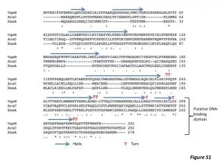
Introduction (cont’) • Transcription factors (TFs) are proteins that regulate gene expression, which serve as integration centers of the different signal-transduction pathways affecting a given gene. • TFs regulate cell development, differentiation, and cell growth by binding to a specific DNA site and regulating gene expression. • The tertiary structures of a large number of TFs are mostly disordered. • Sequence based analysis aimed at identifying the residues in a highly-disordered TF that play key roles in interaction with the DNA is essential for obtaining a comprehensive picture of how the TF functions.
Introduction (cont’) • Two types of binding mechanisms • Sequence-specific (specific) binding • A residue is regarded as involved in sequence-specific binding with the DNA, if one or more heavy atoms in its side-chain fall within 4.5 Å from the nucleobases of the DNA. • Non-specific binding • A residue is regarded as involved in non-specific binding with the DNA, if one or more heavy atoms in its side-chain fall within 4.5 Å from the nucleotide backbone of the DNA.
Specific Binding vs. Non-specific Binding 2PRT:A Red: specific binding residues Blue: non-specific binding residues Purple: both
DNA-binding Mode • Luscombe et al. reported that protein-DNA interactions can be grouped into eight different structural/functional groups • Zinc-coordinating • Zipper-type • Helix-turn-helix (HTH, including “winged” HTH) • Other α-helix • β-sheet • β-hairpin/ribbon • Others • Enzymes Luscombe NM, Austin SE, Berman HM, Thornton JM: An overview of the structures of protein-DNA complexes. Genome Biology 2000, 1(1):reviews001.001 - reviews001.037.
β-sheet, 1DBT Zinc-coordinating, 1A1L Zipper-type, 1YSA HTH, 1BC8
Framework query sequence Sequence-specific binding residue prediction Non-specific binding residue prediction 1st stage Protein-DNA binding mode prediction 2nd stage
Method • Dataset • 253 TF-DNA complexes collected by Chu et al. • Chu WY, Huang YF, Huang CC, Cheng YS, Huang CK, Oyang YJ: ProteDNA: a sequence-based predictor of sequence-specific DNA-binding residues in transcription factors. Nucleic Acids Res 2009, 37(Web Server issue):W396-401. • Classifier • Libsvm package with the Gaussian kernel • http://www.csie.ntu.edu.tw/~cjlin/libsvm/
Feature Set • 1st stage • Evolutionary profile - position specific scoring matrix (PSSM) computed by the PSI-BLAST package • Sliding widow of neighborhood residues information – window size 11 • Labeling: 0: non-binding residues; 1: binding residues • 2nd stage • Predicted non-specific binding residues • 20 amino acids • Secondary structure elements (α-helix, β-sheet, coil) • # of binding residues • Protein chain information • Secondary structure elements (α-helix, β-sheet, coil) • # of total residues in a protein chain • Labeling: zipper-type, helix-turn-helix (HTH), zinc-coordinating, β-hairpin/ribbon, others
Performance Evaluation • In the experiments of the first stage, we repeated the same testing procedure 20 times with randomly and independently generated testing data sets. • The independent testing data set used in each run was derived from 30 TF chains randomly selected from the 253 TF-DNA complexes. • In order to eliminate possible bias present in our collection of TF complexes, we took steps to guarantee that no two TF chains used to generate the testing data set in the same run are homologous with a sequence identity higher than 20%.
Results and Discussion Overall performance
Results and Discussion (cont’) Performance breakdown in terms of secondary structure elements The number of binding residues in β-sheet secondary structure elements is far fewer than the number of binding residues in either a-helix or coil elements. As a result, our proposed method cannot learn sufficient clues in order to identify binding residues in β -sheet elements.
Results and Discussion (cont’) Performance of protein-DNA binding mode prediction The prediction power of sequence-specific binding and non-specific binding residue on β -sheet structure is worse than that of α-helix and coil. The reason we only use non-specific binding residues information as feature set is that non-specific binding residues play a role to stabilize the protein-DNA complex.
Results and Discussion (cont’) Our proposed method is the only predictor listed in this table that has been designed to identify the residues involved in both sequence-specific and non-specific binding with the DNA, while all the other predictors do not distinguish between sequence-specific binding and non-specific binding. It can be easily shown in mathematics that accuracy cannot be higher than sensitivity and specificity simultaneously, which is the case with the numbers reported by Hwang et al. In terms of the F-score, our proposed method is capable of delivering superior performance in comparison with the related works.
1LMB:A Residues colored by red means false positive. Residues colored by blue means false negative. Residues colored by green means true positive. It is obviously that correct binding mode prediction can greatly help the binding residues prediction, especially in difficult case. However, this idea needs more investment to derive a systematic approach.
Modified Framework query sequence Sequence-specific binding residue prediction Non-specific binding residue prediction 1st stage Protein-DNA binding mode prediction 2nd stage
Conclusions • The tertiary structures of a large number of transcription factors are mostly disordered. • It is highly desirable to have a predictor capable of identifying those residues involved in sequence-specific binding and non-specific binding with the DNA. • Our proposed method has been able to deliver • precision 81.70% and 65.47% in sequence-specific and non-specific binding residue prediction respectively • deliver sensitivity 56.85% while combining prediction results of specific binding and non-specific binding. • Concerning a specific type of proteins, a specifically designed predictor should be able to deliver superior performance in comparison with a general-purpose predictor.
DNA Structure nucleotide base nucleotide backbone (sugar phosphate backbone) Image source: doi:10.1093/nar/gkn332
Why 4.5 Å? • The threshold of distance cut-off is based on hydrogen bonding and van der Waals attractions • A hydrogen bond was defined as having a maximum donor–acceptor distance of 3.35 Å and maximum hydrogen–acceptor distance of 2.7 Å. • Atoms were considered to form van der Waals contacts if the distance between them was 3.9 Å and the contact had not been defined as a hydrogen bond
Parameter Selection • 1st stage • Leave-One-Out cross validation • 2nd stage • Leave-One-Out cross validation • Multi-class prediction using one-against-one approach