Introduction and Objective
An Unusual Case of Uterine Fibroid I Adibah, K Norhayati , KC Liew Department of Obstetrics and Gynaecology, School of Medical Sciences, Universiti Sains Malaysia, 16150 Kubang Kerian , Kelantan, Malaysia. Introduction and Objective. The case continues……. The case continues…. Discussion.

Introduction and Objective
E N D
Presentation Transcript
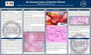
An Unusual Case of Uterine FibroidI Adibah, K Norhayati, KC LiewDepartment of Obstetrics and Gynaecology, School of Medical Sciences, Universiti Sains Malaysia, 16150 KubangKerian, Kelantan, Malaysia Introduction and Objective The case continues…… The case continues….. Discussion Mayer-Rokitansky-Küster-Hauser (MRKH) syndrome is an uncommon congenital anomaly, affecting 1 in 4000 to 5000 of women. It is characterized by vaginal aplasia with normal external genitalia, absence of fibromuscular remnants of the uterus with normally developed fallopian tubes and ovaries. The presence of solid pelvic mass in patients with MRKH is difficult to examine, especially when no vaginal reconstruction has been performed. An imaging technique may provide further information about the origin but may also be misleading. By describing the uncommon association between uterine fibroid and MRKH may make gynaecologist to be aware of its possibility in dealing with pelvic mass in MRKH cases. The patient managed to have a consummative sexual intercourse. However, she had some climacteric symptoms. Physical examination revealed that the secondary sexual characteristics were well developed, and the external genitalia were normal. She had a functional vagina with a length of 4cm achieved through sexual activity, an absence of cervix, and a palpable right pelvic mass. The serum level of oestrogen was normal; however the levels of follicle-stimulating hormone and luteinizing hormone were within menopausal range. The intravenous pyelogram showed duplex right hydroureters with mild hydronephrosis and a single left hydroureter. Transvaginal scan revealed a solid pelvic mass, which could be ovarian in origin. A rudimentary uterus and an atrophic ovary measuring 2cm x 1cm were visualized on the left side. CT scan of the pelvic identified a solid right ovarian mass measuring 5cm x 6cm. The patient underwent a laparotomy. The uterus was substituted by 2 bilateral remnants, of which the one on the right side showed multiple uterine fibroids. The remnants were completely separated. Both fallopian tubes were normal and both ovaries were noted atrophic. Patients with MRKH syndrome have normally developed ovaries and fallopian tubes, and many of them have two uterine remnants. These uterine remnants consist of fibromuscular tissue, which may be the target tissue for ovarian steroids, from which tumours like leiomyomas can originate. A pelvic mass found in MRKH patient needs to be thoroughly evaluated. As illustrated in this case, imaging technique may give some value but may also mislead the physician to the origin of the mass. A better way to evaluate such case is by performing diagnostic laparoscopy, provided that the risk of developing malignancy is low. In conclusion, although the development of uterine fibroid is rare in MRKH patients, it needs to be included in the differential diagnosis of pelvic mass in these patients. Remnants of the uterus Right fallopian tube Fibroid s Right ovary The case References Capraro VJ, Gallego MB. Vaginal agenesis. Am J ObstetGynecol 1976;124:98-107 EfthimiosDeligeoroglou, AntoniosKontoravdis, EvangelosMakrakis, PanagiotisChristopoulos, ApastolosKountouris and George Creatsas. Development of leiomyomas on the uterine mremnants of two women with Mayer-Rokitansky-Küster-Hauser syndrome. FertilSteril 2004;81:1385-1387 Faber M, Stein A, Adashi E. Rokitanski-Küster-Hauser syndrome. ObstetGynecol; 51 (supp 11): 70-3 Marta Lamarca, Ricardo Navario, Maria Eugenia Ballesteros, Salvador Garecia-Agurire, Maria Pilarconte and Jose Antonio Dugue. Leiomyomas in both uterine remnants in a woman with the Mayer-Rokitansky-Küster-Hauser syndrome. FertilSteril 2009;91:931 e13-e15 Metzger DA, Massad LS, Piseitelli JT. Leiomyoma in a mullerian remnant. A case report. J Reprod Med 1988;33:246-8 A 51 year old Malay lady was referred to our centre for further management of primary amenorrhoea. Her previous gynaecologic history included the diagnosis of MRKH made by another service hospital, when she was 18 years of age. She had not undergone any vaginal operation apart from her diagnostic laparoscopy. She was advised to come back to the hospital when she got married which occurred 33 years later. Myomectomy of the uterine fibroids was performed, leaving the uterine remnant to the size of 2cm x 1 cm. The patient was discharged well on the third postoperative day. Histopathology examination of the fibroids confirmed the diagnosis.