Download
1 / 36
430 likes | 621 Views
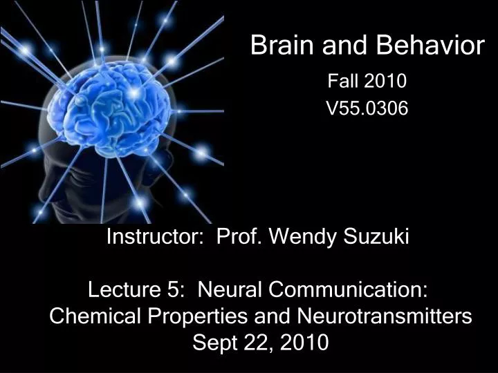
Brain and Behavior Fall 2010 V55.0306. Instructor: Prof. Wendy Suzuki Lecture 5: Neural Communication: Chemical Properties and Neurotransmitters Sept 22, 2010. http://www.hhmi.org/biointeractive/neuroscience/animations.html.

E N D
Brain and Behavior Fall 2010 V55.0306 Instructor: Prof. Wendy Suzuki Lecture 5: Neural Communication: Chemical Properties and Neurotransmitters Sept 22, 2010
http://www.hhmi.org/biointeractive/neuroscience/animations.htmlhttp://www.hhmi.org/biointeractive/neuroscience/animations.html http://www.science.smith.edu/departments/NeuroSci/courses/bio330/squid.html
Two types of communication happen in neurons • Electrical – within the neurons, transmission along the axon • Chemical – between neurons, transmission at the synapse 3
Loewi and “Vagusstoff” • Otto Loewi won Nobel prize for discovery of chemical neurotransmitter acetylcholine in 1936 • 1921 - Stimulation of vagus nerve slowed the heart of a frog. Placing fresh heart into same solution slowed second heart without stimulation. • Deduced that stimulation of vegaus released chemical into solution containing original frog heart. 4
Neurons communicate at synapsesusing neurotransmitters See close up of a synapse on the next slide
What happens when the action potential reaches the axon terminal? Pre-synaptic • AP arrives at axon terminal • 2. Voltage gated Ca2+ channels activated • 3. Ca2+ causes synaptic vesicles to fuse with post-synaptic membrane. • 4. Transmitter molecules released in the synaptic cleft….. Pre-synaptic Post-synaptic 8
What happens when the Neurotransmitter hits the post-synaptic membrane? Post-synaptic 1. The NT’s bind to specific receptors on the post-synaptic membrane 2. This directly or indirect activates ion channels on the post-synaptic membrane 3. Changes in membrane potentials cause EPSP’s or IPSP’s in the post-synaptic cell. Pre-synaptic Post-synaptic 9
Two main types of receptors Ligand-gated receptors – channel is controlled directly by the arrival of the neurotransmitter molecule; “ionotropic” Metabotropic receptors – indirect, “second messenger” process; no direct ion channel activity 10
Receptor Agonists and Antagonists Agonists bind to receptor sites and activate them; drugs that are agonists mimic endogenous ligands Antagonists interfere with receptor activation either by blocking the ligand binding site (competitive) or occupying a remote site (noncompetitive) 11
Same neurotransmitter can interact with different receptor types Receptor type can be different in different locations; either can have excitatory or inhibitory action Excitatory receptors let Na+ in and inhibitory receptors let Cl- in. 12
What keeps the neurotransmitter from just hanging out the in the cleft forever? Answer: Neurotransmitter Housekeeping Pre-synaptic 1. Degradation 2. Re-Uptake Post-synaptic 13
Depolarize – decrease in membrane potential, more positive Hyperpolarize – increase in membrane potential, more negative Threshold – voltage or stimulus intensity that will trigger an action potential Grated Responses 15
Graded potentials: EPSP, IPSP Excitatory postsynaptic potential, positive change Inhibitory postsynaptic potential, negative change 16
Sherrington, on the synapse • Sir Charles Sherrington (1932) Nobel prize winner for work on reflex action, threshold, and summation properties of the synapse • Spatial summation – balance of all excitatory and inhibitory inputs across dendrites and soma • Temporal summation – repeated low-level input sums together to cause an output 17
Neuronal “Information Processing” Spatial Summation vs. Temporal Summation
REVIEW: Two types of communication happen in neurons • Electrical (Action potential)– within the neurons, transmission along the axon • Chemical (Neurotransmission) – between neurons, transmission at the synapse • Electrical (EPSP and IPSP)- in the NEXT neuron 20
Putting it all together: Motor Reflex Speed of transmission: 100 m/s, total time ~40ms 21
Pre-synaptic Post-synaptic 24
What Defines a Neurotransmitter? • Exists in pre-synaptic axon terminals • Pre-synaptic cells contains enzymes for synthesizing substance • Substance is released with AP reach terminals • Specific receptors recognize the substance • Application of the substance produces changes in postsynaptic potential • Blocking release of the substance prevents nerve impulses
Two main types of receptors Ligand-gated receptors – channel is controlled directly by the arrival of the neurotransmitter molecule; “ionotropic” Metabotropic receptors – indirect, “second messenger” process; no direct ion channel activity 27
#1 and #2 transmitters • Glutamate and aspartate are the 2 major excitatory neurotransmitters throughout the brain. Major receptors are AMPA, Kianate and NMDA (All ionotropic) • Gamma aminobyuyric acid (GABA) and Glycineare the major inhibitory transmitters of the brain. 3 kinds of receptors. Gaba A (ionotropic) Gaba B (metabotropic) Gaba C (iontropopic with a choloride channel)
Acetylcholine distribution: Memory Cholinergic cells in basal forebrain and dorsal brainstem send axons to cerebral cortex, hippocampus, amygdala, cerebellum. Present in PNS, neuromuscular junction. Widespread degeneration in Alzheimer’s disease Role in learning, memory, Arousal Receptors: Nicotinic: ionotropic Muscarinic: G-protein coupled 31
Dopamine distribution: Reward Dopaminergic cells in VTA and substantia nigra send to 2 main projections: limbic system, hippocampus, and cortex; basal ganglia including N. Accumbens. Abnormalities in mesolimbic pathway associated with schizophrenia; degeneration in mesostriatal pathway in Parkinson’s disease. Role in motor control, learning, reward mechanisms. Receptors: D1-D5 receptors of various characteristics. 32
Norepinephrine distribution: Arousal Noradrenergic cells in Locus Coeruleus in pons and Lateral Tegmental Area in the midbrain send axons throughout the coretx, subcortex, cerebellum, and spinal cord. Also important action in PNS. Role in overall regulation of mood and arousal. Note: epinephrine is also called adrenelin Receptors: Alpha 1 and 2, Beta, 1 and 2 all metabotropic receptors. 33
Serotonin distribution: Sleep, Waking, Mood* Serotonergic cells in Raphe nuclei of midbrain and brainstem send axons widely in CNS. Affect limbic system and basal ganglia, regulate sleep centers in brainstem. Role in sleep and waking, anxiety, mood regulation. Receptors: Many different kinds! 34
Opiates: pain/pleasure Opiate receptors are found throughout the brain: Limbic system (emotion and motivated behavior), hippocampus (learning and memory), thalamus (sensory relay nucleus) Endogenous ligands (endorphins) have been identified; mediate pain relief; Exogenous ligands (morphine, heroin) bind to opiate receptors Receptors: Delta, kappa and mu are all G-protein couples d metabotropic recptors Opiate receptor distribution in rat brain 35