Download

1 / 42
460 likes | 709 Views
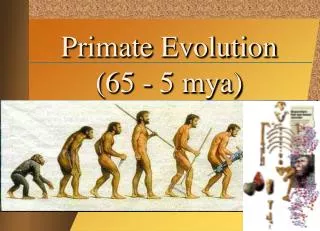
The Lower Extremity: Functional Consequences of Bipedality Form Follows Function. (From J.G. Fleagle’s Primate Adaptation & Evolution , 1988). Bipedal Locomotion ONLY in humans!!!. (From R.M. Alexander’s The Human Machine , 1992). Why are we so unique? MUST STAND UPRIGHT!.

E N D
The Lower Extremity: Functional Consequences of Bipedality Form Follows Function (From J.G. Fleagle’s Primate Adaptation & Evolution, 1988)
Bipedal Locomotion ONLY in humans!!! (From R.M. Alexander’s The Human Machine, 1992) Why are we so unique? MUST STAND UPRIGHT!
4 Design Considerations for Bipedal Gait and Upright Posture 1) Stability in Upright Posture 2) Ability to Raise & Control Trunk Over Hindlimbs 3) Ability to Balance on One Leg 4) Walk with Feet Underneath Body
Stability lower extremities larger & heavier than upper extremities Weebles wobble but they don’t fall down!
Ability to Raise & Control Trunk Over Hindlimbs Gluteus Maximus sacral attachment
Tm TW W Ability to Balance on One Leg Well-developed Hip Abductors gluteus medius gluteus minimus
ANGLE OF FEMUR • 14-15 degrees • moves CM more directly over base of support • DON’T HAVE TO SHIFT LATERALLY WHEN YOU WALK!
ilium sacrum head acetabulum neck ischium pubis Obturator foramen lesser trochanter greater trochanter ANTERIOR VIEW POSTERIOR VIEW
Comparison to Shoulder • the hip is a “weight bearing” joint • both are ball-and-socket joints • acetabulum much deeper than glenoid fossa • both have a “labrum” to increase depth of the socket • hip has more bony support than shoulder • left and right shoulder girdles are more independent than the corresponding portions of the pelvis/femur
Gender • Females have pelvic girdles that are lighter, thinner and wider than their male counterparts. • The female pelvis flares out more laterally in the front and the sacrum is wider in the back, creating a broader pelvic cavity than males.
Pelvic movement • Concomitant movement of the pelvic girdle and the thigh at the hip joint are necessary for efficient joint actions. • Movements of the pelvis are described by monitoring the ilium - specifically the anterior superior iliac spine.
Anterior Tilt • forward tilting and downward movement of the pelvis • occurs when the hip extends
Posterior Tilt • tilting of the pelvis posteriorly • occurs when the hip flexes
Lateral Tilt • tilting of the pelvis from neutral position to the right or left • lateral tilt tends to occur naturally when you support your weight on your leg • this allows you raise your opposite leg enough to swing through during gait
Pelvic Rotation • rotation of the pelvis defined by the direction in which the anterior aspect of the pelvis moves • occurs naturally during unilateral leg movements (walking) • as the right leg swings forward during gait the pelvis rotates left
Hip Joint capitis femoris ligament (round ligament) acetabular labrum • The femoral head and acetabulum have large amounts of spongy, trabecular bone to help attenuate forces. • Approximately 70% of the head of the femur articulates with the acetabulum. sagittal view of right hip
Hip Ligaments anterior view of right hip iliofemoral (Y-shaped) Resists extension, internal rotation and some external rotation. pubofemoral ligament Resists abduction and some external rotation.
Hip Ligaments ischiofemoral ligament Resists adduction and internal rotation. Note: none of these ligaments restrict flexion. posterior view of right hip
Femoral Neck • The neck holds the femur away from the pelvis. • It is formed by cancellous trabecular bone and reinforced with cortical bone, particularly on the inferior portion. • The angle of inclination is measured in the frontal plane and typically ranges from 90 to 135 degrees.
Coxa Vara • If the angle of inclination is less than 125 degrees it is termed coxa vara. • This shortens the limb, increases the effectiveness of the abductors, reduces the load on the femoral head and increases the load on the femoral neck.
Coxa Valga • If the angle of inclination is greater than 125 degrees it is termed coxa valga. • This lengthens the limb, reduces the effectiveness of the abductors, increases the load on the femoral head and reduces the load on the femoral neck.
Angle of Anteversion • The angle of the femoral neck in the transverse plane is termed the angle of anteversion. • Normally the femoral neck is rotated anteriorly 12 to 14 degrees with respect to the femur.
Excessive Anteversion • Excessive anteversion beyond 14 degrees causes the head of the femur become uncovered. • In order to keep the head of the femur within the acetabulum a person must internally rotate the femur.
Retroversion • If the angle of anteversion is reversed so that it moves posteriorly, it is termed retroversion. • This condition causes the person to externally rotate the femur.
Hip Range of Motion Movement Range flexion 70-140o hyperextension 4-15o adduction 20o abduction 30o internal rotation 70o external rotation 90o
Primary Hip Flexors psoas major iliacus (aka iliopsoas)
Assisting Hip Flexors: pectineus rectus femoris sartorius tensor fascia latae
Assisting Hip Flexors: pectineus tensor fascia latae sartorius rectus femoris
Hip Extensors Gluteus maximus Hamstrings biceps femoris semitendinosus semimembranosus
semimembranosus semitendinosus biceps femoris M T B lateral medial
long head short head Biceps Femoris
Hip Extensor Hip Abductors gluteus maximus gluteus medius & minimus
Hip Adductors pectineus adductor brevis adductor longus adductor magnus anterior view
Hip Adductors gracilis posterior view
Medial Rotation of the Hip • due primarily to the gluteus medius and minimus • extension of hip tends to laterally rotate femur so medial rotators needed to neutralize this effect • not usually performed against resistance, thus not a lot of muscular support • medial much weaker than lateral rotation Assisting Muscles semimembranosus, semitendinosus, tensor fascia latae, and hip adductors
Lateral Rotation of the Hip • lateral rotation - 5 muscles • lateral rotation is a natural movement in human gait to accommodate pelvic rotation
Muscle Activity During Walking Muscle Footstrike Midsupport Toe-off Swing Decel. Dorsiflexors ******* ** ** Intrinsic Foot *** Gluteus Maximus * ****** Gluteus Medius ******* * Gluteus Minimus ** ***** * Hamstrings ******* * ** Iliopsoas *** Plantar Flexors * ** Quadriceps * ***** * Sartorius ** * Tensor Fascia Latae * ** * *** Thigh Adductors **** * ** *
Hip Fractures • occurs in neck of femur • usually due to a decreased bone mineral density • 87% are 65 or older • current annual cost is more than $9.8 billion • accounts for more hospital days, by far, than any other musculoskeletal injury • results in increased mortality, reduced mobility, and, for many, the inability to live independently • American Academy of Orthopaedic Surgeons
Hamstring Injuries • few activities require simultaneous hip flexion and knee extension • usually little hamstring stretch except for specific exercises • hamstrings susceptible to strain due to this poor extensibility • injuries most often occur during sprinting - particularly when muscle is fatigued
Hamstring Injuries - Theories • overstretching of muscle • for example: during overstriding • development of maximal tension when muscle is fully elongated • development of max tension necessary to act antagonistically to quads which are stronger
opposite same hurt leg W hurt leg W Which side of the body do you use a cane on when your hip is hurt?