Download
1 / 52
520 likes | 729 Views
Hearing. Functions of the ear. Hearing (Parts involved): External ear Middle ear Internal ear Equilibrium sense (Parts involved): Internal ear. Anatomical consideration. Outer ear: Pinna External canal Tympanic Membrane (funnel shaped, pointing inward). Anatomical consideration.

E N D
Functions of the ear • Hearing (Parts involved): • External ear • Middle ear • Internal ear • Equilibrium sense (Parts involved): • Internal ear
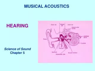
Anatomical consideration • Outer ear: • Pinna • External canal • Tympanic Membrane (funnel shaped, pointing inward)
Anatomical consideration • Middle ear: • Air filled cavity • Three bones: • Mallaus • Incus • Stapes (with its foot sitting on the oval window of the inner ear)
Anatomical consideration • Inner ear: • Bony and membranous labyrinth
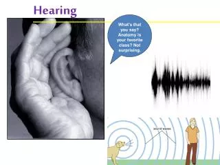
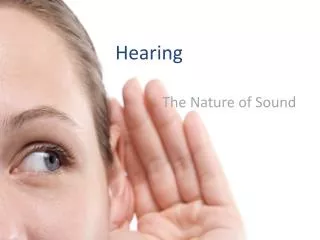
Nature of Sound • Sound is produced from alternate compression and rarefaction of air molecules by vibrating body
Characteristics of sound • 1- Pitch (Tone) depend on No. of cycle/sec. Human ear can detect sound waves with freq.. 20-20000 cycle /sec • 2- Intensity (Loudness) depend on amplitude • 3- Quality depend on the over tone or interference
Functions of the ear • External ear: • Act as funnel to collect sound • Sound localisation (front, back, high, low) • Protection
Functions of the ear • Middle ear: it is a space between tympanic membrane and the inner ear (opens via Eustachian tube into nasopharynx) • Content: • 1- Air • 2- Ossicles • Malleus • Incus • Stapes • 3- Muscles • 1- Tensor tympani • 2- Stepedius
Functions of the middle ear • 1- Ossicles: • Manbrium of the malleus attached to the back of the tympanic membrane and its short process attached to the incus. • The incus then articulates with the head of the stapes, and its foot plate attached to the oval window
Functions of the middle ear • 2- Muscles: • Muscles contract reflexly in response to loud sound (over 70dB) • Contraction of the tensor tympani pulls the manubruim & makes the tympanic m. tens. Thus decreasing the vibration. • Contraction of the stapeduis pull the foot plate outward so that vibration are reduced • (protection from constant loud noise, but not sudden noise, latency of 40-80 msec.
Transmission of sound through the middle ear • sound waves vibrate the tympanic m. • Tympanic m moves the handle of malleus • Incus moves • Stapes move in & out of the oval window. The pressure transmitted through cochlea cause stimulation of hair cells in the organ of corti, which will stimulate the auditory nerve
Middle ear magnifying effect • 1- The force from a large surface area (Tympanic m.) are concentrated to a small (oval window) the ratio is 17=1 • 2- Lever action of ossicles = the lever action of ossicles increase the force of movement 1.3 times • ▲ the total increase 17 X 1.3 = 22 times
Inner ear • Anatomy: • Cochlea (snail like, coiled tubular system laying deep in the temporal bone) • Bony labyrinth • Membranous labyrinth
Cochlea • It is a system of three coiled tubes through its length • The basilar m. & the reissners m divide it into three canals: • Scala Vestibuli • Scala Media • Scala Tympani
Composition • Scala Vestibuli: Na high K low • Scala Tympani: Na high K low • Scala Media : Na low K high
Organ of Corti • Located (resting) on the basilar m. • Contain inner & outer hair cells • Extend from base to apex
Hair cells • Steroclia extend from the top • Arrangement: • Three rows of outer hair cells (attached to the reticular lamina or tectorial m.) • One row of inner hair cells (not attached to tectorial m.)
Function of inner hair cells • Striocellia not embedded in tectorial m. but bent by fluid movement under the tectorial m. • They are primary receptors for sound, transducing fluid movement in cochlea into action potential in the auditory nerve
Function of the outer hair cells • Large number, but stimulate only small fraction of nerve fibres in the cochlear nerve • If damaged, significant loss of hearing (they control the sensitivity of inner hair cells to particular sound frequency)
Receptors & Endocochlear potentials • Sound transmission into the inner ear cause upper & lower movements of the reticular m. (tectorial m.) • »»»»» produce bending of steriocillia of the hair cells alternatively open & close cation channels at the tip of the steriocillia
»»»»» (inward current) depolarization »»»»» (outward current) hyperpolarisation • »»»»» the net results is depolarization
Production of cells receptors potentials • »»»»» release of neurotransmitter • »»»»» production of action potentials
The Central Auditory pathway • This pathway begins in the organ of corti • End in the primary auditory cortex (are 41& 42, superior temporal gyrus in the temporal lobe of the brain) • Fibres end in the auditory area, where it is heard, then interpretation occurs in the auditory association areas (wernikes area)
The Central Auditory pathway • There is a bilateral cortical connection of auditory area • Thus damage to one side only slightly reduces hearing
Sound localization • Differences in the time arrival of the sound wave at the ears (time-lag) • Differences in the loudness
Masking effect • Presence of background noise affect the ability to hear another sound, due to some receptors are in refractory period • Masking is more clear if two sound are having the same frequencies
Noise pollution is an environmental hazard • Exposure to sound intensity above 80dB may damage outer hair cells
Conduction of sound wave • Air conduction: • Normal situation of hearing, sound travel in air causes vibration of Tympanic m., transmitted by ossicles to the oval window
Conduction of sound wave • Bone conduction: • Sound cause vibration of skull bones directly transmitting the sound vibration to the cochlea (eg when placing tuning fork on the head or mastoid process)
Deafness • Conductive deafness • Perceptive deafness
Conductive deafness • Impairment of sound transmission through external or middle ear due to: • Wax • Repeated infection • Perforated drum • Destruction of ossicles • Osteosclerosis (pathological fixation of stapes on the oval window)
Conductive deafness • All sound frequencies are equally affected • Bone conduction is better than air conduction
Perceptive deafness • Due to congenital or damage to cochlea or auditory nerve pathway due to: • Toxins (antibiotics, gentamycine) • Inflammation • Vascular • Tumour Both air and bone conduction are affected
Test of hearing • Audiometer • Weber test • Rinnes test