Exploring the Appendicular Skeleton: Limbs & Girdles
670 likes | 704 Views
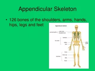
Dive into the anatomy of the appendicular skeleton encompassing pectoral & pelvic girdles, upper limb bones, and their articulations. Discover the detailed structures like scapula, humerus, radius, and ulna, ending with the intricate hand bones.

Exploring the Appendicular Skeleton: Limbs & Girdles
E N D
Presentation Transcript
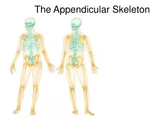
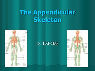
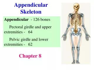
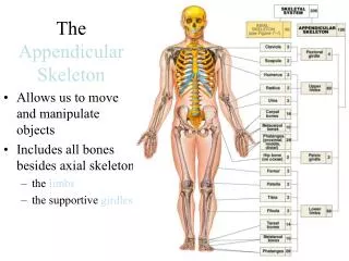
8 The Appendicular Skeleton
Repition….Repition….Repition…. Repition….Repition….
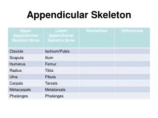
I. The Appendicular Skeleton A. Pectoral girdle B. Pelvic girdle C. Upper and lower limbs
II. The Pectoral Girdle A. clavicle B. scapula - Provides attachment for many muscles that move the upper limb - Girdle is very light and upper limbs are mobile
Acromio-clavicularjoint Clavicle Scapula Articulated pectoral girdle
III. Clavicle A. Extends horizontally across the superior thorax 1. sternal end articulates with the manubrium 2. acromial end articulates with scapula ► Provides attachment for muscles ► Holds the scapulae and arms laterally ► Transmits compression forces - upper limbs
Sternal (medial)end Posterior Anterior Acromial (lateral)end Right clavicle, superior view
IV. Scapula A. Lie on the dorsal surface of the rib cage B. Located between ribs 2–7 C. Have three borders 1. superior 2. medial (vertebral) 3. Llateral (axillary) D. Have three angles 1. Lateral 2. superior 3. inferior
Superior border Suprascapularnotch Acromion Superior angle Coracoidprocess Glenoidcavity Subscapularfossa Lateral border Medial border Inferior angle Right scapula, anterior aspect
Coracoid process Suprascapular notch Superior angle Acromion Supraspinousfossa Glenoidcavityat lateralangle Spine Infraspinousfossa Lateral border Medial border Right scapula, posterior aspect
V. The Upper Limb A. 30 bones form each upper limb B. Grouped into bones of the: 1. Arm 2. Forearm 3. Hand
VI. Arm Humerus ► The only bone of the arm ► Longest and strongest bone of the upper limb ► Articulates with the scapula at the shoulder ► Articulates with the radius and ulna at the elbow
Greatertubercle Head ofhumerus Lessertubercle Deltoid tuberosity Coronoidfossa Medial epicondyle Capitulum Trochlea Anterior view
Head ofhumerus Greatertubercle Deltoidtuberosity Olecranonfossa Medial epicondyle Lateralepicondyle Trochlea Posterior view
The humerus of the right arm and detailed views of articulation at the elbow (anterior). Coronoidfossa Humerus Medialepicondyle Capitulum Trochlea Head ofradius Coronoidprocess ofulna Radialtuberosity Radial notch Radius Ulna Anterior view at the elbow region
The humerus of the right arm and detailed views of articulation at the elbow (posterior). Humerus Olecranonfossa Olecranon Lateralepicondyle Medialepicondyle Head Neck Ulna Radius Posterior view of extended elbow
VII. Forearm A. radius & ulna articulate with each other B. The interosseous membrane 1. Interconnects radius and ulna C. radius is lateral - ulna is medial
VIII. Ulna Major landmarks of the ulna ► Olecranon ► Radial notch ► Trochlear notch ► Coronoid process ► Ulnar styloid process
Olecranon Troclearnotch Head Neck Coronoid process Interosseousmembrane Ulna Radius Head of ulna Ulnar styloidprocess Radial styloidprocess Anterior view
Olecranon Trochlear notch View Coronoid process Radial notch Proximal portion of ulna, lateral view
View Proximal portion of ulna, lateral view
IX. Radius Major landmarks of the radius: ► Head ► Neck ► Radial tuberosity ► Styloid process
The humerus of the right arm and detailed views of articulation at the elbow. Coronoidfossa Humerus Medialepicondyle Capitulum Trochlea Head ofradius Coronoidprocess ofulna Radialtuberosity Radial notch Radius Ulna Anterior view at the elbow region
The humerus of the right arm and detailed views of articulation at the elbow. Anterior view at the elbow region
X. Hand Includes the following bones ► Carpals—wrist ► Metacarpals—palm ► Phalanges—fingers
XI. Carpus (Wrist) Carpal bones A. Proximal row from lateral to medial Scaphoid, lunate, triquetral, and pisiform B. Distal row from lateral to medial Trapezium, trapezoid, capitate, and hamate thumb► “Sally left the party thumb►to take Carmen home”
Carpal bones thumb► “Sally left the party thumb►to take Carmen home” IV III II V Hamate I Capitate Trapezium Trapezoid Pisiform Scaphoid Triquetrum Lunate Ulna Radius Anterior view of right hand
Carpal bones IV III II V I Anterior view of right hand
XI. Metacarpals Metacarpals form the palm ► Numbered I - V : beginning with the pollex (thumb)
IV III II V I Anterior view of right hand
XII. Phalanges ► Numbered I - V : beginning with the pollex (thumb) ► Except for the thumb, each finger has three phalanges Proximal, middle, and distal
Phalanges Distal Middle Proximal Distal Proximal Anterior view of right hand
XIII. Pelvic Girdle A. Attaches lower limbs to the spine B. Supports visceral organs C. Attaches to the axial skeleton by strong ligaments D. Acetabulum is a deep cup that holds the head of the femur E. Paired coxal bones (hip bones) and the sacrum
XIV. Pelvic Girdle A. Three separate bones (fuse together) ► Ilium ► Ischium ► pubis
Iliac crest Iliac fossa Anterior superioriliac spine Ilium Coxal bone Anteriorinferior iliacspine (os coxaeor hip bone) Sacrum Coccyx Pubis Acetabulum Ischium Pubic symphysis Pelvic girdle Pubic arch
Iliac fossa Sacrum Coccyx Pelvic girdle Pubic arch
Ilium Ala Iliac crest Anteriorsuperioriliac spine Posteriorsuperioriliac spine Anterior inferioriliac spine Posterior inferioriliac spine Acetabulum Greater sciaticnotch Ischial spine Lesser sciaticnotch Pubis Ischium Ischialtuberosity Ilium Ischium Pubis Lateral view, right hip bone
Ala Ilium Ischium Pubis Lateral view, right hip bone
XV. The Lower Limb A. carries the entire weight of the erect body B. lower limb are thicker and stronger than those of upper limb C. Divided into three segments 1. thigh 2. leg 3. foot
XVI.Thigh A. Femur—the single bone of the thigh ► longest and strongest bone of the body ► head of femurarticulates with the acetabulum
Foveacapitis Neck Greatertrochanter Head Lesser trochanter Gluteal tuberosity Lateralcondyle Lateralepicondyle Medial condyle Lateralepicondyle Medialepicondyle Anterior view Posterior view Femur (thigh bone)
Anterior view Posterior view Femur (thigh bone)