Download

1 / 59
660 likes | 1.51k Views
Objectives. At the end of this segment, when given a clinical presentation, gross specimen, and/or photomicrograph students will be able to:Compare and contrast the clinical presentations, etiologies, pathogenesis, and gross and microscopic changes found in developmental, inflammatory, circulatory,

E N D
1. Pathology of the Small Intestine Aiman Zaher, MD
2. Objectives At the end of this segment, when given a clinical presentation, gross specimen, and/or photomicrograph students will be able to:
Compare and contrast the clinical presentations, etiologies, pathogenesis, and gross and microscopic changes found in developmental, inflammatory, circulatory, mechanical, and neoplastic disorders of the small intestine.
3. Objectives At the end of this segment, when given a clinical presentation, gross specimen, and/or photomicrograph students will be able to:
Predict the clinical complications associated with diseases of the small intestine.
Define the words in the glossary
4. Glossary Adhesions
Ileus
Intussusception
Volvulus
5. Structure and Function
6. Gross
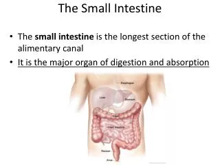
~6 meters long
Duodenum (retroperitoneal), jejunum, ileum
Blood supply
Blood supply to duodenum
Superior pancreaticoduodenal artery (branch of gastroduodenal artery).
Inferior pancreaticoduodenal artery (branch of superior mesenteric artery).
Blood supply to remainder of small intestine is from superior mesenteric artery
7. The purpose of the small intestine is for terminal digestion & absorption of foodstuffs.
Histology
Lined with many villi
Villi have three cell types:
Columnar absorptive cells with microvilli
Mucin-secreting goblet cells
Endocrine cells
Between the villi are the crypts of Lieberk�hn
Contain stem cells, goblet cells, endrocrine cells, and Paneth cells (contain antimicrobial proteins)
Duodenum contains numerous submucosal glands, called Brunner�s glands
8. Congenital Anomalies
9. Congenital Anomalies Heterotopia
Usually pancreas, but can be gastric mucosa appearing as small nodules in the mucosa or intestinal wall
Atresia and Stenosis
Duodenal atresia is most common, followed by jejunum and ileum
Stenosis can also be acquired e.g. intussusceptions
10. Congenital Anomalies Meckel Diverticulum
Failure of the vitelline duct (connects the developing gut to the yolk sac) to involute
Found on the anti-mesenteric side of gut within two feet of ileocecal valve
Contains all three layers of normal bowel wall (true diverticulum)
Heterotopic rests of gastric or pancreatic tissue found in 50% ? peptic ulceration ? bleeding
Complications include intussusception, incarceration or perforation, but most are incidental findings
12. Enterocolitis
13. Diarrhea & Dysentery Diarrhea
An increase in stool mass, frequency or fluidity in most patients
Characterized by pain, urgency, perianal discomfort and incontinence
Dysentery
Low-volume, painful, bloody diarrhea
14. Diarrhea & Dysentery - Mechanisms Secretory diarrhea
Passage of >500 ml/day of watery stools, isotonic with plasma
e.g. rotavirus, E. coli, V. cholaerae, villus adenomas and excessive laxative use
Osmotic diarrhea
Passage of > 500 ml/day of stools, osmolality exceeds that of plasma by > 50 mOsm.
e.g. lactase deficiency and antacids
Exudative diseases
Passage of frequent purulent, bloody stools
e.g. Shigella, Salmonella
15. Malabsorption
bulky stools with excess fat that floats on the water (steatorrhea) and increased osmolality
e.g. Celiac Sprue and Crohn disease
Deranged Motility
Improper gut neuromuscular function ? variable patterns of increased stool volume
e.g. surgical reduction of bowel length, diverticula Diarrhea & Dysentery - Mechanisms
16. Infectious Enterocolitis Intestinal diseases of microbial origin
Characterized by diarrhea and in some instances ulceration of the bowel
Causes >12,000 deaths per day among children in developing countries and equals � of all deaths before age 5 worldwide
17. Infectious Enterocolitis Viruses
Acute, self-limited infectious diarrhea is most frequently caused by enteric viruses.
Rotavirus � outbreaks in infants
Norwalk viruses � outbreaks in school children and adults; Norwalk virus is responsible for the majority of cases of nonbacterial food-borne epidemic gastroenteritis in all age groups.
Adenoviruses � outbreaks in infants
Astroviruses � outbreaks in children
18. Infectious Enterocolitis Bacteria
E. coli (food borne; invasive & non-invasive forms)
Vibrio cholerae (water borne; enterotoxin ? secretory diarrhea)
S. aureus (food poisoning; preformed toxin)
Salmonella and Shigella (invasive bloody diarrhea; toxins)
MAI (AIDs associated)
Clostridium difficile (antibiotic associated colitis)
Parasites
e.g. Giardia lamblia, Entamoeba histolytica
19. Miscellaneous Intestinal Inflammatory Disorders AIDS
Diarrheal illness in 50% of AIDS patients in developed countries
Some malbsorption, some ulcerative colitis, infections with other organisms; possibly due to HIV mucosal damage, itself
Complications of Transplantation (particularly bone marrow)
Pre-transplant: Blunted villi, degeneration and flattening of crypt cells with decreased mitosis due to direct toxic injury
Graft versus host: focal crypt cell necrosis: severe, watery diarrhea
Drug-induced intestinal injury
Focal ulceration when a pill sticks to the mucosa or enterocolitis (most commonly NSAIDs)
Radiation
endothelial cell injury ? ischemic fibrosis & stricture
Acute radiation enteritis: anorexia, cramping, and malabsorption
Chronic radiation enteritis: inflammatory enteritis
Neutropenic colitis (typhlitis)
20. Malabsorption Syndromes
21. Malabsorption Syndromes Malabsorption - Definition
Characterized by suboptimal absorption of fats, fat-soluble and other vitamins, proteins, carbohydrates, electrolytes and minerals, and water
22. Pathogenesis - Malabsorption Syndromes Defective Intraluminal Digestion
Pancreatic insufficiency
Zollinger-Ellison Syndrome
Bacterial overgrowth
Primary Mucosal Cell Abnormalities
Defective terminal digestion (lactose intolerance)
Defective epithelial transport (abetalipproteinemia)
Reduced Small Intestinal Surface Area
Crohn Disease
Celiac Sprue
Lymphatic Obstruction
TB
Lymphoma
Infection
Whipple disease
Tropical Sprue
Iatrogenic
Gastrectomy
Distal ileal resection
23. Malabsorption Syndromes Clinical Presentation
Chronic diarrhea and steatorrhea:
Pass bulky, frothy, greasy, yellow, or gray stools
weight loss, anorexia and abdominal pain
In US pancreatic insufficiency, Celiac Sprue and Crohn disease are most important
Multiple systems involved; if prolonged leads to:
Anemia, petechiae, hemorrhages, dermatitis, bone pain, peripheral neuropathy, latent tetany, menstrual and reproductive disturbances, among other symptoms
Symptoms due to vitamin deficiencies
24. Celiac Disease AKA Celiac Sprue and Gluten-Sensitive enteropathy
Definition
Chronic disease with characteristic mucosal lesion of the small intestine and impaired nutrient absorption that improves on withdrawal of wheat gliadins and related grain proteins from diet
Epidemiology
Almost exclusively Caucasians
25. Celiac Disease Etiology
Hypersensitivity to wheat gluten and gliadin associated with HLA-DQ2 and DQ8
Pathogenesis
T-cell-mediated hypersensitivity
26. Celiac Disease Morphology
Grossly, mucosa appears flat or scalloped, or even normal
Microscopically, diffuse enteritis with marked atrophy or total loss of villi
Epithelial cells degenerated with loss of microvilli and increased intraepithelial lymphocytes
Crypts exhibit increased mitotic activity.
Morphology mimics other diseases, like tropical sprue
Mucosa will revert back to normal when stimulus taken away.
29. Celiac Disease Clinical Features
Diarrhea & failure to thrive in infants, but adults might not present with malabsorption syndromes till their 50s
Anti-gliadin or �anti-endomysial� antibodies favors diagnosis
Definitive diagnosis requires
Clinical documentation of malabsorption
Small bowel biopsy results
Improvement of symptoms upon gluten withdrawal
Clinical Complications
Risk of neoplasia e.g. non-Hodgkin lymphoma, small intestinal adenocarcinoma, and esophageal SCC (50-100X risk)
30. Tropical Sprue (Postinfectious Sprue) Celiac-like malabsorption syndrome, seen in people of the tropics or visiting the tropics, including the Caribbean.
No specific causal agent found, but enterotoxigenic organisms implicated
Responds to antibiotic therapy
Changes similar to those of Celiac disease, but is seen at all levels of the small intestine and not associated with lymphoma
31. Whipple Disease A rare systemic disease of primarily the intestines, joints, and CNS, caused by gram-positive actinomycete, Tropheryma whippelii
Pathogenesis unknown
Patients are usually white, M:F = 10:1, 40-50 years of age
Lamina propria is laden with distended macrophages, containing tiny, rod-shaped bacilli that are PAS positive
33. Whipple Disease Clinical
Presents with malabsorption syndrome, sometimes of years� duration
Arthropathy is often the initial presentation
Lymphadenopathy & hyperpigmentation >50%
Also, polyarthritis, cardiac, and neurologic signs and symptoms
Responds to broad spectrum antibiotics
34. Disaccharidase (Lactase) Deficiency Disaccharidase is an apical membrane enzyme that cleaves lactose.
35. Pathogenesis
Incomplete breakdown of disaccharide (lactose) into glucose and galactose
Leads to osmotic diarrhea
Bacterial fermentation of unabsorbed sugar ? increased hydrogen production and gaseous symptoms Disaccharidase (Lactase) Deficiency
36. Disaccharidase (Lactase) Deficiency Congenital form
Presents in infants on exposure to milk or milk products
Explosive, watery diarrhea and abdominal distension that stops when taken off milk
Acquired form
More common
Adults, blacks & native americans > whites; sometimes related to viral or bacterial enteric infection
No morphologic changes
37. Abetalipoproteinemia Deficiency of betalipoprotein that is required for intestinal transport of chylomicrons
Chylomicrons Chylomicrons
38. Circulatory Disorders
39. Ischemic Bowel Disease General
Can be restricted to either the small or large intestine, or both
Infarctions seen with acute occlusion of celiac, superior and inferior mesenteric arteries
Insidious loss of one vessel may go unnoticed due to rich anastomoses
Etiology
Arterial thrombosis
Arterial embolism
Venous thrombosis
Nonocclusive ischemia; e.g. cardiac failure, shock, etc.
Miscellaneous
Radiation injury
Volvulus
Stricture
40. Ischemic Bowel Disease Types of lesions
Transmural Infarction
All layers due to sudden occlusion of major vessels
Bowel swollen, gangrenous and perforates in few days
Mural & Mucosal Infarction
Most commonly due to hypoperfusion in watershed areas
Necrosis of mucosa only; mucosa hemorrhagic; serosa normal
Chronic Ischemia
Mucosal atrophy; ulcerations; mural fibrosis
Can lead to stricture
44. Ischemic Bowel Disease Clinical Features
Uncommon, but grave. 50-75% death rate
Short time between symptoms and perforation
Transmural infarcts
sudden severe abdominal pain and tenderness; sometimes nausea, vomiting and bloody diarrhea or melena
shock and vascular collapse in hours
peristalsis is diminished
Mucosal and mural infarcts
may not be fatal if cause corrected
nonspecific abdominal complaints and intermittent bloody diarrhea, but may progress to extensive infarction & sepsis
Chronic ischemic infarcts
insidious with intermittent bloody diarrhea, resembling inflammatory bowel disease
45. Obstructions/ Dilatations
46. Hernias Etiology
Usually weakness in wall of peritoneal cavity may permit protrusion of a pouch-like, serosa-lined sac of peritoneum
Most common sites
inguinal and femoral canals
umbilicus
surgical scars
Clinical Significance
Segments of viscera protrude and become trapped e.g. small bowel ? Ischemia
Incarceration = permanent trapping of bowel loop due to edema from impaired venous drainage
Strangulation = compromised arterial supply & venous drainage ? infarction
48. Adhesions Etiology
Inflammation (peritonitis) e.g. surgery, infection, radiation and endometriosis
As healing occurs, get adhesions between bowel loops, bowel wall, & surgical site
Complications
Twisting of bowel loops around peritoneal fibrous bands, strangulating & obstructing the bowel
51. Intussusception Etiology
One segment of bowel, constricted by a wave of peristalsis, telescopes into another more distal segment
Once in peristalsis, wedges in further
Mesentery pulled in, and ischemia ensues
Pathogenesis
Infants � usually no underlying cause, but can be associated with rotavirus infection
Adults � usually an intralumenal mass or neoplasm
53. Volvulus Definition
Complete twisting of a bowel loop about its mesenteric base.
Produces obstruction & infarction
Most often occurs in large redundant loops of sigmoid colon and small intestine.
55. Neoplasms of the Small Intestines 3-6% of GI Tumors
Adenomas and mesenchymal tumors most frequent benign tumors
Malignant tumors rare �Only ~1% of GI tumors
e.g. adenocarcinomas and carcinoids followed by lymphomas and sarcomas
56. Adenomas Epidemiology
25% of benign tumors
Most in Ampulla of Vater region
Higher incidence in patients with familial polyposis
Clinical
Occult blood in stool
Rarely, obstruction and intussuseption
Morphology
Tumors resemble those seen in colon
Those that extend into ampular orifice render themselves difficult to remove surgically short of pancreatoduodenectomy to remove entire ampullary region
57. Adenocarcinoma Epidemiology
Age - 40-70 years
Majority in duodenum
Major risk factor is inflammation from CD
Clinical Features
Weight loss, cramping, nausea, vomiting
Obstructive jaundice if located in Ampulla
Fatigue if blood loss
Clinical Complications
Most neoplasms ? penetrates wall ? invade mesentery ? metastasized to regional nodes ? ? liver by time diagnosis is made
70% survival at 5 years with surgery
59. References Kumar, Abbas, and Fausto: ROBBINS AND COTRAN PATHOLOGIC BASIS OF DISEASE, 7th Edition, pp.828-870.