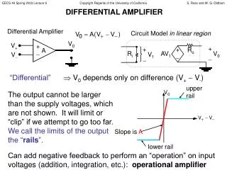
Differential Centrifugation
Differential Centrifugation. By Sophie Legg. Differential Centrifugation. This is the most common method of fractionating cells Fractionation is the separation of the different organelles within the cell. Method:.


Differential Centrifugation
E N D
Presentation Transcript
Differential Centrifugation By Sophie Legg
Differential Centrifugation • This is the most common method of fractionating cells • Fractionation is the separation of the different organelles within the cell
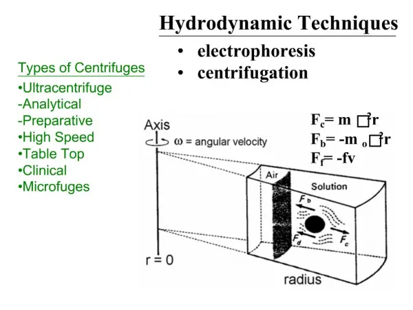
Method: • 1. Cut tissue in an ice-cold isotonic buffer. It is cold to stop enzyme reactions, isotonic to stop osmosis and a buffer to stop pH changes. • 2. Grind tissue in a blender to break open cells. • Filter to remove insoluble tissue
4. Centrifuge filtrate at low speeds ( 1000 X g for 10mins ) • This pellets the nuclei as this is the densest organelle
5. Centrifuge at medium speeds ( 10 000 x g for 30 mins ) • This pellets mitchondria which are the second densest organelle
6. Centrifuge at high speeds ( 100 000 x g for 30 mins) • This pellets ER, golgi apparatus and other membrane fragments
7 Centrifuge at very high speeds ( 300 000 x g for 3hrs) • This pellets ribosomes
Investigating Cell Function • Differential Centrifugation allows us to look at each organelle within the cell • We can look at the individual organelles and study them in detail • This helps to determine each organelles function within the cell
The Electron Microscope • Microscopes allow us to see living organisms which are too small to be seen by the naked eye • The electron microscope uses beams of electrons rather than light to illuminate the specimen • A beam of electrons has an effective wavelength of less than 1 nm so it can be used to resolve small sub-cellular ultra-structure • The development of the electron microscope allowed biologists to view the organelles within a cell for the first time
The transmission microscope. (TEM) Works like a light microscope, it transmits a beam of electrons through a thin specimen Then focussing the electrons to form an image on a screen This is the most common form of electron microscope and gives good resolution. The scanning electron microscope (SEM) This scans a fine beam of electron onto specimen and collects electrons scattered by surface This has poor resolution but gives good 3-D images There are two types of electron microscope
Disadvantages of the Electron Microscope • The specimens must be fixed in plastice and viewed in a vacuum and so they must be dead • Sometimes specimens can be damaged by the electron beam and must be stained with an electron-dense chemical