Download
1 / 31
320 likes | 622 Views
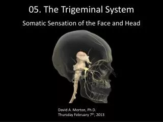
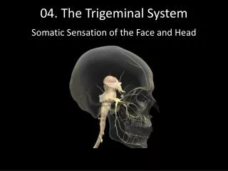
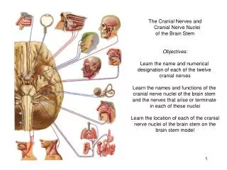
The Cranial Nerve Nuclei and the Trigeminal System. General Organization of the Cranial Nerve Nuclei and the Trigeminal System. Introduction to the cranial nerves and nuclei A. The 12 cranial nerves, their location, and functions.

E N D
General Organization of the Cranial Nerve Nuclei and the Trigeminal System • Introduction to the cranial nerves and nuclei A. The 12 cranial nerves, their location, and functions. B. Categories of cranial nerves, and the columnar organization of their nuclei. II. Functional anatomy of the trigeminal system A. Ascending pathway for touch (using main trigeminal sensory nucleus). B. Ascending pathway for pain and temp (using spinal trigeminal nucleus).
General Organization of the Cranial Nerve Nuclei and the Trigeminal System III. Regional anatomy A. Key components of the trigeminal system within the medulla (e.g., spinal trigeminal nucleus + tract). B. Components within the pons – main trigeminal accessory nucleus. C. Midbrain: mesencephalic trigeminal nucleus. D. VPM + projections to 1° somatic sensory cortex.
I. Introduction to the Cranial Nervesand Nuclei A. 12 pairs: • Olfactory – enters telencephalon (cerebral hemispheres). • Optic – a part of the diencephalon. Both are purely sensory. III. Oculomotor IV. Trochlear – the only cranial nerve on dorsal brainstem surface. Both are purely motor and exit from midbrain.
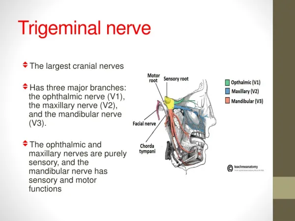
I. Introduction to the Cranial Nervesand Nuclei 12 pairs: V. Trigeminal – in middle of pons – and have mixed sensory/motor with separate roots. VI. Abducens – motor VII. Facial – mixed – muscles of facial expression and taste. VIII. Vestibulocochlear – sensory – vestibular and auditory. All originate at the ponto-medullary junction
I. Introduction to the Cranial Nervesand Nuclei 12 pairs: IX. Glossopharyngeal – mixed (main = sensory; post 1/3 of tongue for taste + pharynx). X. Vagus – mixed – many functions: taste, visceral sensory (i.e., gut, aortic arch receptors, peripheral autonomic (gut, respiratory, cardiac), some muscles (pharnyx, larynx, lungs). XI. Spinal accessory – motor (neck muscles). XII. Hypoglossal – motor (tongue muscles).
Mnemonics • “On Old Olympus’ Towering Top, A Finn And German Viewed A Hop” Or more contemporarily: • “Oh, Oh, Oh To Take A Family Vacation! Go Vegas After Hours!” To remember which are sensory and/or motor: “Some Say Marry Money But My Brother Says Better Brains Matter More”
Cranial nerves are analogous to spinal nerves in many ways (as the bs is analogous to sc), serving sensory and motor functions of the head, rather than trunk and limbs. 1 Difference: a greater variety of sensory modalities served - “special senses”, such as sight and hearing.
B. Seven functional categories of cranial nerves and the columnar organization of their nuclei. • General somatic afferents [somatic sensations of head (touch, pain, proprioception)]. • General visceral afferents [visceral sensations, chemoreception]. • General somatic motor (inervation of striatal muscle]. • General visceral motor [axons of autonomic neurons]. • Spinal somatic afferents [vision, hearing, balance]. • Spinal visceral afferents [taste, smell]. • Special visceral motor [muscles from bronchial arches].
Cranial nerve nuclei: Sensory, motor – analogous to dorsal, ventral horn neurons of sc. Organization of each category of nuclei in rostral-caudal columns. See Figs. 6-3 and 6-4
Columnar Organization of Cranial Nerve Nuclei Fig. 6-3 Note that sensory columns are located lateral to motor columns.
General somatic motor extraocular and tongue muscles. • Spinal visceral motor jaw, facial, throat, and many neck muscles (derived from the broncial arch). • General visceral motor pre-ganglionic autonomics. • Visceral afferents (general and spinal combined): • Solitary Nucleus: rostral special – taste…VII, IX, X. caudal general – sometimes from visceral organs and chemoreceptors…IX, X. • Special somatic afferents - vestibular, cochlear nuclei…VIII. • General somatic afferents – trigeminal nuclei…V.
II. Functional Anatomy of the Trigeminal System • Serving somatic sensations of head. • Analogous to dorsal column – medial lemniscal and anterolateral systems of the lower body (separate pathways mediate touch and pain-temp). • Ascending pathway for touch (using main trigeminal sensory nucleus). The most important cn in these systems is V (VII, IX, X cover the small area shown in Fig. 6-6 – Shows the ascending pathways. Review for yourself that touch is mediated by larger-diameter myelinated axons.
Most neurons synapse in the main decussate in the pons. Trigeminal sensory nucleus ascend in trigeminal lemniscus (near medial lemniscus) VPM (note the other system used VPL) 1° sensory cortex (specific area within the homonculus – lateral) (Fig. 6-6A). A smaller ipsilateral (non-decussating) pathway processes mechanical stimuli from teeth and soft tissues of the oral cavity. B. Ascending pathway for pain/temp (using spinal trigeminal nucleus). * review diam, unmyelinated fibres enter at pons and descend to the various levels (divisions) of the spinal trigeminal nucleus.
Fig. 6-6A. Ascending Trigeminal Pathways for Touch: main trigeminal sensory nucleus
Fig. 6-6B. Trigeminal pathway for Pain and Temperature: spinal trigeminal nucleus 3 Components: oral, interpolar, and caudal nuclei. After synapsing, ascending pathway is crossed and travels in trigeminalthalamic tract of the anterolateral system. This path is important mainly for facial and dental pain. Pathway synapses onto VM past central gyrus for discriminative aspect of pain and also interlaminar nuclei diffuse projections for emotional aspects of pain (should also be familiar).
III. Regional Anatomy of the Trigeminal System • Cranial nerve involvement and key components within medulla 3 trigeminal sensory roots, all serving different regions of the head: - opthalmic (skin, mucous membranes) - maxillary (skin, mucous membranes) - mandibular (includes most of mouth interior, except for pharynx and post 1/3 of tongue). Fig. 6-8. Note: very little overlap compared to spinal dermatomes significant regarding lesions. Contributions from VII, IX, X: skin of ear (VII, X), larynx (X), pharynx, post 1/3 of tongue (IX), dura mater (V, X).
Fig. 6-8. Somatotopic Organization:
Peripheral sensory ganglia: V – semilunar IX and X – superior VII – geniculate Specifically for stretch receptors of jaw muscles: Soma contained in brainstem rather than in a ganglion. 3rd trigeminal nucleus: mesencephalic trigeminal nucleus. Somatotropic organization of the ascending trigeminal pathways: slices organized like an ‘inverted face’ (Fig. 6-8).
Rostral-caudal somatotropic organization of medullary trigeminal spinal nuclei (like an onion skin) (Fig. 6-8B). May correspond to caudal, interpolar and oral divisions.
Caudal Medullary Level (Fig. 6-9) Spinal trigemenal nuc: Organized much like sc dorsal horn within the caudal medulla ‘Rexed’s Laminae’ Spinal trigeminal tracts – much like a continuation of Lissauer’s Tract. Reticular formation is continuous with intermediate zone.
Components within the Pons – main trigemenal sensory nucleus subserving touch (tactile sensations of face, head, teeth). This nucleus, along with the trigeminal lemniscus, can be viewed in a pontine section: Fig. 6-12. This is the trigeminal equivalent of dorsal column nuclei. Note: dorsal 1/3 of nucleus receives mechanoreceptive signals from teeth and mouth and gives rise to ipsilateral pathway (dorsal). Therefore, VPM receives bilateral projections for each region.
Midbrain: mesencephalic trigeminal nucleus. • As noted earlier, this nucleus is equivalent to a ganglion, containing the axons of jaw, muscle stretch receptors. • Found in midbrain, in lateral portions of periaquaductal grey (PAG). • A projection to trig motor n. mediates the jaw jerk reflex (analogous to knee-jerk reflex). • In this section, can also see trig lemniscus (med to med lem).
D. Thalamus (VPM) and projections to 1° somatic sensory cortex. Fig. 6-14: VPL: somatic sensations from trunk and limbs. VPM: somatic sensations from head (medial magnocellular). VPM: somatic sensations from taste relay nuc. (parvocellular) Projections to 1° somatic sensory cortex (face area, Fig. 6-15). Fig. 6-14
Fig. 6-15: “Face Area” of Primary Somatic Sensory Cortex: