Download
1 / 78
820 likes | 1.44k Views
Cranial nerves and their nuclei. 鄭海倫 整理. Cranial Nerves. Figure 13.4a. Location of the cranial nerves. Anterior cranial fossa: C.N. 1–2 Middle cranial fossa: C.N. 3-6 Posterior cranial fossa: C.N. 7-12. Functional components in nerves. General Somatic Efferent

E N D
Cranial Nerves Figure 13.4a
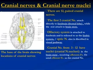
Location of the cranial nerves • Anterior cranial fossa: C.N. 1–2 • Middle cranial fossa: C.N. 3-6 • Posterior cranial fossa: C.N. 7-12
Functional components in nerves • GeneralSomatic Efferent • SpecialVisceral Afferent • GSEGSAGVEGVA • (SSE) SSASVE SVA
The floor of the 4th ventricle in the embryonic rhombencephalon
Sp: special sensoryB:branchial motor Ss: somatic sensorySm: somataic motor Vi: visceral sensory A: preganglionic autonomic (visceral motor)
STT: spinothalamic tract • CST: corticospinal tract • ML: medial lemniscus
Sensory nerve • Olfactory (1) • Optic (2) • Vestibulocochlear (8)
Motor nerve • Oculomotor (3) • Trochlear (4) • Abducens (6) • Accessory (11) • Hypoglossal (12)
Mixed nerve • Trigeminal (5) • Facial (7) • Glossopharyngeal (9) • Vagus (10)
Innervation of branchial muscles • Trigemial • Facial • Glossopharyngeal • Vagus
Cranial Nerve I: Olfactory Table 13.2(I)
Cranial Nerve II: Optic • Arises from the retina of the eye • Optic nerves pass through the optic canals and converge at the optic chiasm • They continue to the thalamus (lateral geniculate body) where they synapse • From there, the optic radiation fibers run to the visual cortex (area 17) • Functions solely by carrying afferent impulses for vision
Cranial Nerve II: Optic Table 13.2(II)
Cranial Nerve III: Oculomotor • Fibers extend from the ventral midbrain, pass through the superior orbital fissure, and go to the extrinsic eye muscles • Functions in raising the eyelid, directing the eyeball, constricting the iris, and controlling lens shape
Cranial Nerve III: Oculomotor Table 13.2(III)
1.Oculomotor nucleus(GSE) • Motor to ocular muscles: rectus (superior對側, inferior同側and medial同側),inferior oblique同側, levator palpebrae superioris雙側 2. Edinger-Westphal nucleus(GVE) • to ciliaryganglion ciliarlis and sphincter pupillae muscles
Oculomotor nucleus: a series of cell columns or subnuclei M: medial longitudinal fasciculus PAG: periaqueductal gray
Cranial Nerve IV: Trochlear • Fibers emerge from the dorsal midbrain and enter the orbits via the superior orbital fissures; innervate the superior oblique muscle • Primarily a motor nerve that directs the eyeball
Cranial Nerve IV: Trochlear Table 13.2(IV)
Trochlear nucleus (GSE) • To contralateral (對側) superior oblique muscle • Located at the level of the inferior colliculus • It indents the medial longitudinal fasciculus
Cranial Nerve VI: Abducens • Fibers leave the inferior pons and enter the orbit via the superior orbital fissure • Primarily a motor nerve innervating the lateral rectus muscle Table 13.2(VI)
Abducens nucleus (GSE) • To lateral rectus muscle • Located in the caudal pons beneath the floor of the 4th ventricle
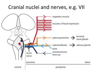
ICP: inferior cerebellar peduncle ML: medial lemniscus SpVt: spinal trigeminal tract VII: facial nerve VIIg: internal genu of the facial nerve VIIn: facial motor nucleus
DMN X: dorsal motor nucleus of the vagus ST: solitary tract Sol: nucleus of the solitary tract 4V: 4th ventricle
Cranial Nerve V: Trigeminal • Composed of three divisions: ophthalmic (V1), maxillary (V2), and mandibular (V3) • Fibers run from the face to the pons via the superior orbital fissure (V1), the foramen rotundum (V2), and the foramen ovale (V3) • Conveys sensory impulses from various areas of the face (V1) and (V2), and supplies motor fibers (V3) for mastication
Cranial Nerve V: Trigeminal Table 13.2(V)
1. Main sensory nucleus 2. Nucleus of the spinal trigeminal: receives information of pain and temperature 3. Mesencephalic nucleus Central processes motor nuclei of trigeminal Peripheral processes mandibular division 4. Trigeminal motor nucleus (SVE): inervates muscles of mastication
SCP: superior cerebellar peduncle MCP: middle cerebellar peduncle V: trigeminal nerve
P: pyramid FC: fasciculus cuneatus NC: nucleus cuneatus
Cranial Nerve VII: Facial • Fibers leave the pons, travel through the internal acoustic meatus, and emerge through the stylomastoid foramen to the lateral aspect of the face • Mixed nerve with five major branches • Motor functions include facial expression, and the transmittal of autonomic impulses to lacrimal and salivary glands • Sensory function is taste from the anterior two-thirds of the tongue
Cranial Nerve VII: Facial Table 13.2(VII)