Download
1 / 26
260 likes | 630 Views
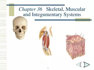
Chapter 36 Skeletal, Muscular and Integumentary Systems. Section Outline. Section 36-1. I. The Skeletal System A. The Skeleton: two subdivisions: Axial and Appendicular 1. Functions a. supports the body

E N D
Section Outline Section 36-1 • I. The Skeletal System • A. The Skeleton: two subdivisions: Axial and Appendicular • 1. Functions • a. supports the body • b. protects internal organs (brain and internal organs • c. provides for movement (levers for muscles to act on) • d. stores minerals (calcium) • e. site for red blood cell production (produced in red marrow) 2
Skull Clavicle Sternum Scapula Humerus Ribs Radius Vertebral column Pelvis Ulna Carpals Metacarpals Phalanges Femur Patella Fibula Tibia Tarsals Metatarsals Phalanges The Skeletal System Section 36-1 Axial Skeleton Axial Skeleton Appendicular Skeleton 3
B. Structure of Bones - a solid network of living cells and protein fibers, surrounded by calcium mineral salts • 1. periosteum – a tough layer of connective tissue • surrounding a bone • 2. Haversian canals – a network of tubes running • through compact bone that carry blood vessels • 3. spongy bone – strong but light bone found and the • ends of long bones, and between compact bone in ribs and skull bones • 4. bone cells- three types • a. osteocytes – mature bone cells that maintain • bone structure • b. osteblasts – create new bone tissue • c. osteoclasts – break down bone Section Outline Section 36-1 4
Spongy bone Haversian canal Compact bone Compact bone Periosteum Bone marrow Spongybone Osteocyte Artery Periosteum Vein Figure 36-3 The Structure of Bone 5
Section Outline Section 36-1 • Development of Bones • 1. embryonic skeletons are composed of cartilage • 2. cartilage is replaced by bone during embryonic • development • 3. the process of replacement is called ossificaton • 4. in early adulthood all the cartilage is replaced by • bone (ossification) 6
D. Types of Joints 1. Immovable Joints – allow no movement – skull bones 2. Slightly Movable Joints – allow some movement – between bones of lower leg, and between vertebrae 3. Freely Movable Joints – movement in one or more directions a. ball and socket joint –movement in many directions (shoulder and hip) b. hinge joint – movement in one direction (knee and elbow) c. pivot joint – one bone rotates around another (lower arm) d. saddle joint – bones slide in two directions (between wrist and hand) Section 36-1 7
Clavicle Humerus Ball-and-socket joint Radius Pivot joint Scapula Ulna Humerus Femur Patella Fibula Metacarpals Hinge joint Carpals Saddle joint Tibia Figure 36-4 Freely Movable Joints and Their Movements Ball-and-Socket Joint Pivot Joint Hinge Joint Saddle Joint 8
E. Structure of joints and skeletal disorders • 1. ends of bones in freely moveable joints are covered • with cartilage for protection • 2. the joint is surrounded by a tough joint capsule that • has two layers • a. ligaments are strips of connective tissue that hold bones together and stabilize the joint • b. the other layer produces synovial fluid, which lubricates the joint • 3. bursa are small sacs found in a joint that reduce friction and act as cushions • 4. Skeletal System Disorders • a. bursitis – inflammation of the bursa • b. osteoporosis – a loss of calcium from bones which causes them to weaken Section Outline Section 36-1 9
Muscle Tendon Femur Patella Bursa Ligament Synovial fluid Cartilage Fat Fibula Tibia Figure 36-5 Knee Joint Section 36-1 10
Section Outline Section 36-2 II. Muscular System– causes movement of body parts and materials A. Types of muscle tissue 1. skeletal – connected to the skeletal system a. striations – alternating light and dark bands seen under a microscope b. under voluntary nervous control c. have many nuclei in each cell 2. smooth muscle – located in internal organs a. no striations present b. involuntary nervous control 3. cardiac muscle – found in the heart a. striations present b. involuntary nervous control 11
Section Outline Section 36-2 B. Muscle Contraction 1.muscle cells are made up of 2 fibers called myofibrils a. a thin filament called actin b. a thick filament called myosin 2. a muscle contraction happens when actin and myosin slide past each other 3. actin and myosin form cross bridges between them which allows a contraction to take place 4. ATP is used to power the reactions that cause contraction C. Control of Muscle contractions 1.skeletal muscle contraction is controlled by the nervous system 2. a nerve ending connects to each muscle cell at a neuromuscular junction 12
Section Outline Section 36-2 3.when a nervous impulse is sent down a nerve, a chemical is released that causes contraction to begin a. acetylcholine is the neurotransmitter that is released D. Muscle and Bone interaction 1.muscles can only contract, so they are arranged in opposing pairs 2. muscles connect to bones by connective tissue strips called tendons 13
Cycle Diagram 1 Myosin forms cross-bridge with actin 5 2 Myosin returns to original shape Cross-bridge changes shape 4 3 Cross-bridge releases actic Actin pulled 14
Figure 36-7 Skeletal Muscle Structure Section 36-2 15
Figure 36-8 Muscle Contraction Section 36-2 Relaxed Muscle Z line Myosin Actin Z line Movement of Actin Filament Actin Cross-bridge Sarcomore Binding sites Myosin Contracted Muscle Cross-bridges Z line 16
Figure 36-8 Muscle Contraction Section 36-2 Relaxed Muscle Z line Myosin Actin Z line Movement of Actin Filament Actin Cross-bridge Sarcomore Binding sites Myosin Contracted Muscle During muscle contraction, the knoblike head of a myosin filament attaches to a binding site on actin, forming a cross-bridge. Cross-bridges Z line 16
Figure 36-8 Muscle Contraction Section 36-2 Relaxed Muscle Z line Myosin Actin Z line Movement of Actin Filament Actin Cross-bridge Sarcomore Binding sites Myosin Contracted Muscle During muscle contraction, the knoblike head of a myosin filament attaches to a binding site on actin, forming a cross-bridge. Powered by ATP, the myosin cross-bridge changes shape and pulls the actin filament toward the center of the sarcomere. Cross-bridges Z line 16
Figure 36-8 Muscle Contraction Section 36-2 Relaxed Muscle Z line Myosin Actin Z line Movement of Actin Filament Actin Cross-bridge Sarcomore Binding sites Myosin Contracted Muscle During muscle contraction, the knoblike head of a myosin filament attaches to a binding site on actin, forming a cross-bridge. Powered by ATP, the myosin cross-bridge changes shape and pulls the actin filament toward the center of the sarcomere. The cross-bridge is broken, the myosin binds to another site on the actin filament, and the cycle begins again. Cross-bridges Z line 16
Figure 36-11 Opposing Muscle Pairs Opposing Muscle Pairs Movement Movement Biceps (relaxed) Biceps (contracted) Triceps (relaxed) Triceps (relaxed) 17
Section 36-3 III. Integumentary System: Skin, hair, nails and several glands A. Functions 1. a barrier against infection and injury 2. helps regulate body temperature 3. removes waste products 4. provides protection from ultraviolet light 18
B. Two main layers: Epidermis and Dermis • 1. epidermis – upper layer of skin • a. top layer of skin is dead, and made up of a • waterproofing protein called keratin • b. deeper layer is living and produces a dark • pigment called melanin (UV protection) • c. the epidermis is avascular (no blood vessels) • 2. dermis – contains blood vessels, nerve endings, hair • follicles, glands, and smooth muscle • a. blood vessels help to regulate heat loss and gain • b. glands produce sweat and oils to cool the body • and keep the skin flexible and waterproof Section 36-3 19
Regulator of body temperature Protector against UV radiation Remover of waste products Barrier to infection Dermis Epidermis Inner layer Outer layer Concept Map Section 36-3 Skin functions as a is made up of the which is the which is the 20
Figure 36-13 The Structure of Skin Section 36-3 21
Video 1 5
Video 2 Video 2 5