Download
1 / 23
240 likes | 457 Views
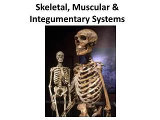
Integumentary and Skeletal Systems. Test Notes. Skeletal System. The skeletal system consists of : B ones Cartilage Tendons Ligaments

E N D
Integumentary and Skeletal Systems Test Notes
Skeletal System • The skeletal system consists of: • Bones • Cartilage • Tendons • Ligaments • Bonemaintains the shape of the body, protects internal organs, is a lever system for muscles to act upon, and is a site of mineral storage and blood-cell formation. • Cartilage forms a fetal model of bone, covers the ends of bones, and provides a firm, flexible support. • Tendons attach muscle to bone. • Ligaments attach bone to bone. • To help you remember: LLL, TTT (ligaments are like to like, tendons are two types)
Skeletal System (Tendons and Ligaments) tendon • extracellular matrix of tendons and ligaments is made up of primarily collagen fibers which makes them very tough like ropes or cables. ligament
Skeletal System (Cartilage) hyaline • Three types of cartilage: • Hyaline • Elastic • Fibrocartilage • Hyaline cartilage is most intimately associated with bone function and development elastic fibrocartilage
Skeletal System (Bone) cancellous • Bone Tissue Types: • Cancellous (Spongy) • Compact • Cancellous Bone: • Located mainly in the epiphyses of long bones & the interior of all other bones. • Consists of a lacy network of bone with many small, marrow-filled spaces. • Compact Bone: • Mostly solid matrix and cells. • Forms most of the diaphysis of long bones & the thinner surfaces of all other bones. compact
Skeletal System (Bone) • Bone is composed of an organic matrix (mostly collagen) that provides flexible strength and an inorganic matrix (hydroxyapatite) that provides compressional strength (weight bearing). • Hydroxyapatite are calcium phosphate crystals • Bone is formed in thin sheets of extracellular matrix called lamellae • Compact bone consists of cells called osteocytes located within spaces in the matrix called lacunae
Skeletal System (Bone) • Ossification is the formation of bone by osteoblasts • Intramembranous ossification occurs when osteoblasts begin to produce bone in connective tissue membranes (primarily in bones of the skull) • Endochondralossification is the process that produces most of the skeleton, occurs when bones develop from cartilage models, and occurs when osteoblasts invade the spaces left by dying cartilage cells
Skeletal System (Bone) • Four types of bone, based on their shape: • Long • Short • Flat • Irregular
Skeletal System (Bone Shapes) Long bones are longer than they are wide (bones of upper & lower limbs). Short bones are approximately as broad as they are long (bones of ankle & wrist). Flat bones have a relatively thin flattened shape (some skull bones, ribs, & sternum). Irregular bones do not fit in the other shape categories (vertebrae & facial bones). Long bones Short bones Flat bones Irregular bones
Epiphysis • Diaphysis • Hyaline (Articular) Cartilage • Periosteum BONE STRUCTURE - Long Bone
Skeletal System (Bone) • a long bone has a diaphysis (shaft) and an epiphysis (each end) • the epiphyseal plate (growth plate), is the site of growth in length of a long bone, and is found between each epiphysis and the diaphysis • a long bone has a medullary cavity (filled with yellow marrow) in the diaphysis, has cancellous bone at the ends (filled with red marrow) • has an endosteum lining the medullary cavity • outer surface covered by a connective tissue layer- periosteum
Structure of a Long Bone Figure 6.3a-c
Medullary Cavity – hollow chamber filled with bone marrow Red Marrow (blood) Yellow Marrow (fat) Endosteum – lining of the medullary Inside the Long Bone
Compact (wall of the diaphysis) Spongy (cancellous, epiphysis) Types of Bone Tissue
Review the Structure of a Long Bone Matching quiz at http://www.mhhe.com/biosci/ap/holehaap/student/olc2/chap07matching01.html
MATRIX - where the bone cells live OSTEOCYTES - mature bone cells, enclosed in tiny chambers called LACUNAE OSTEOCYTES form rings (LAMELLAE) around a HAVERSIAN CANAL which houses blood vessels Osteocytes are linked by CANALICULI Haversian Canals are linked by VOLKMAN's CANALS Microscopic Structure
BONE DEVELOPMENT & GROWTH • Intramembranous bones – flat, skull • Endochondral bones – all other • ALL BONES START AS HYALINE CARTILAGE, areas graduallly turn to bone • PRIMARY OSSIFICATION CENTER (shaft) • SECONDARY OSSIFICATION CENTER (ends)
EPIPHYSEAL DISK (growth plate) is a band of cartilage between the epiphysis and diaphysis These areas increase bone length as the cells ossify Cartilage becomes OSTEOBLASTS become OSTEOCYTES Bone Development & Growth
RESORPTION OSTEOCLASTS - dissolve bone tissue to release minerals, process is called RESORPTION