Interactive Digestion Quiz: Explore Comparative Anatomy of Mandibles and Teeth
Test your knowledge of animal digestion and oral anatomy with this engaging quiz developed by Sorcha McCaughley and Mark Brims, and approved by experts Gawain Hammond and Maureen Bain. Explore the structures of the jaws, teeth, and gastrointestinal tract through interactive questions, visual aids, and comparative anatomy for species including canines, felines, and equines. Supported by The Chancellor’s Fund, this educational tool is perfect for students of veterinary science and animal biology.

Interactive Digestion Quiz: Explore Comparative Anatomy of Mandibles and Teeth
E N D
Presentation Transcript
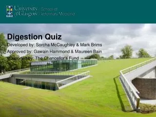
Digestion Quiz Developed by: Sorcha McCaughley & Mark Brims Approved by: Gawain Hammond & Maureen Bain Supported by: The Chancellor’s Fund
Digestion Quiz START! Developed by: Sorcha McCaughley & Mark Brims Supported by: The Chancellor’s Fund
Choose a Region… • The jaws and teeth • Oesophagus • Abdominal Gastro-Intestinal Tract (Note: See Respiration for Hyoid bones)
I want to view the…. • Mandible and Maxilla • Teeth • Comparitive Mandibles & TMJ’s • Comparitive Teeth
Comparative Mandible & TMJ’s Bovine Mandible Feline Mandible Coronoid Condylar Angular TMJ Feline Skull, Lateral Equine Skull, Lateral Page 2 >
Comparative Mandible/Maxilla Feline Maxilla Feline Mandible Vomer Palatine Fissure Symphysis Choose a Region < Page 1
Comparative Teeth 1 Ovine Skull, Lateral Equine Mandible & Maxilla, D/V Ruminant Dental Formula: 0-0-3-3 3-1-3-3 Feline Skull, Lateral Equine Dental Formula: 3-1-3(4)-3 3-1-3-3 Page 2 > Feline Dental Formula: 3-1-3-1 3-1-2-1
Comparative Teeth 2 Equine Maxilla, Lateral Here it is seen that the roots of the equine cheek teeth are embedded in the Maxillary Sinus. The sinus gets larger as the horse ages and the teeth continually erupt. This arrangement can cause problems as dental disease may pass through the sinus into the respiratory tract. By entering the sinus surgically the roots of the teeth may be accessed and the infected tooth can be removed. Maxillary Sinus Cheek tooth roots <Page 1 Choose a Region
Mandible & Maxilla Part 2 • What is structure A? • Body of Mandible • Coronoid Process • Condylar Process • What is structure B? • Coronoid Process • Angular Process • Condylar Process • What is structure C? • Coronoid Process • Angular Process • Condylar Process • Do you know what the orange line D represents? • Answer Canine Mandible, Lateral A B C Canine Mandible, Dorso-Ventral D
Mandibular Symphysis • This is the Mandibular Symphysis. It is the joining point between the two halves of the Mandible. It may appear more fused in older animals. • Back to Choose a Region
Incorrect • No, this is not the body of the mandible. • The body is the horizontal part extending rostrally: Try Again!
Incorrect • No, this is not the Condylar Process. • Here is an example of the Condylar Process: Try Again!
Correct! • Yes! A is the Coronoid Process of the mandible. • Here is another example: • Try part B
Incorrect • No, this is not the Coronoid Process. • Here is an example of the Coronoid Process: Try Again!
Incorrect • No, this is not the Angular Process. • Here is an example of the Angular Process: Try Again!
Correct! • Yes! B is the Condylar Process of the mandible. • Here is another example: • Try part C
Correct! • Yes! C is the Angular Process of the mandible. • Here is another example: • Try part D
Mandible & Maxilla Part 1 • a) Is the top radiograph of a Mandible or Maxilla? • Mandible • Maxilla • b) Do you know which joint is shown in the blue circle on the bottom radiograph? • Answer
Incorrect • No, this is not the Mandible! It forms the bottom part of the jaws. • Here is the Mandible: Try Again!
Correct! • Yes! This is the Maxilla. It can be recognised by the presence of the Palatine Fissure andVomer: • Try part b)
Temporo-Mandibular Joint This is the Temporo-Mandibular Joint. It is the joint connecting the Mandible to the Maxilla and the rest of the skull. It is formed by the Condylar Process and the Mandibular Fossa of the Skull. Here is another example: Now try Mandible & Maxilla part 2
Teeth • a) Can you identify the roots, pulp cavity, dentine & enamel on the top radiograph? • Answers • b) On the bottom radiograph, which teeth are incisors, which are canines and which are premolars & molars? • What is the complete dental formula of the dog? • Answers
Teeth Pulp Cavity Enamel Dentine Root Now try part b)
Teeth Canine (1) Premolars (4) Incisors (3) Molars (3) Complete dental formula: 3-1-4-2 3-1-4-3 Back to Choose a Region
Oesophagus • Which is the Oesophagus, A or B? • A • B B A
Correct! Yes! B is the Oesophagus! It lies dorsal to the Trachea in the neck. It is not normally easy to see on radiographs as it is not rigid and is collapsed: In the example, it has had contrast introduced. Back to Choose a Region (6 is a small volume of gas in the oesophagus. The rest of its length cannot be seen)
Incorrect No! A is the Trachea. It is black on radiographs as it is rigid and gas filled. It lies ventral to the Oesophagus. Here is another example: Try Again! (6 is a small volume of gas in the oesophagus. The rest of its length cannot be seen)
Abdominal GIT I want to view the: • Liver • Spleen • Stomach and Duodenum • Jejunum and Ileum • Caecum, Colon and Rectum Choose a Region
Liver 1 • a) The position of the Liver has been highlighted in the top radiograph by injecting contrast into the venous system. What limits the extent of the liver cranially (red arrows)? • Lungs • Stomach • Diaphragm • b) Contrast has been introduced into certain blood vessels on the bottom radiograph. Which vessels are shown? • Answers
Liver Veins Portal Vein Portal Vein Abdominal Veins (Mesenteric etc.) The radiograph shows the abdominal veins supplying the Portal Vein. The second radiograph, above, shows how the lobes of the liver are heavily vascularised by the branches of the Portal Vein. Now try Liver 2
Incorrect No, the lungs themselves do not limit the extent of the Liver. They are located in the thoracic cavity, they do not come into direct contact with the Liver in the Abdominal Cavity. Try Again!
Incorrect No, the stomach does not limit the cranial extent of the Liver. The stomach lies caudal to the liver and may affect its extent in that direction. Try Again!
Correct! • Yes! The red arrows point to the diaphragm. The liver lies pressed against the diaphragm and takes on its shape in situ. • This can be seen in this feline example where air has entered the peritoneum and shows the outline of the liver: Try part b)
Liver 2 • The bile duct of this liver has been injected with contrast. What is A? • Gall Bladder • Spleen • Stomach A
Correct! • Yes! A is the Gall Bladder. It fills with bile collected from the lobes of the liver. It sits between the Quadrate and Right Medial lobes. • Here is an example in an isolated liver: • Can you name the other lobes? Back to Abdominal GIT
Incorrect No! A is not the Spleen. The Spleen is not connected to the Bile duct, is larger and located more caudally than A. Here is an example (labeled): Try Again!
Incorrect No! A is not the Stomach. The Stomach is not connected to the Bile duct, is larger and located more caudally than A. Here is an example (with contrast): Try Again!
Spleen • Do you know where in the abdomen the spleen is located? • Answer
Spleen location Here is the location of the spleen. It is located on the left side of the abdomen and lies along the greater curvature of the stomach. As it is connected to the stomach by the gastrosplenic ligament, its exact position depends on the stomach. Back to Abdominal GIT page
Stomach & Duodenum Canine Stomach & Duodenum with contrast • a) Can you name the regions of the stomach labelled A, B, C & D? • What types of glands are present in these regions? • Answers • b) Where is the Duodenum? What side of the abdomen is it on? What are its regions? • Answers Right D C A B Cd. Cr. Left Click here for lateral and Feline views of the stomach
Lateral & Feline Stomach Canine Stomach & Duodenum with contrast Feline Stomach Cd. Cr. Feline Stomach Back to Stomach & Duodenum
Regions of the Stomach A = Cardia (Oesophagus enters. Has mucous secreting Cardiac glands) B = Fundus (Has HCl secreting Parietal cells and pepsin secreting Chief cells. Also endocrine cells secreting Gastrin) C = Body (Has HCl secreting Parietal cells and pepsin secreting Chief cells. Also endocrine cells secreting Gastrin) D = Pylorus (Has mucous cells and endocrine glands which secrete Gastrin. Duodenum begins here) Pylorus Body Cardia Fundus Cd. Cr. Try part b)
The Duodenum Canine Stomach & Duodenum with contrast Right Here is the Duodenum. It leaves the stomach on the right side of the abdomen, descends caudally, turns back on itself, then ascends cranially a short distance. After this, it becomes the Jejunum. Descending Duodenum Cranial Flexure Ascending Duodenum Caudal Flexure Left Back to GIT page
Jejunum & Ileum • The radiograph shows the loops of Jejunum in the circled area. The Ileum is also located in this area but cannot be distinguished from the Jejunum. • Some of the loops have a black appearance, why is this? Is this normal? • Answer
Jejunum & Ileum Answer • The Jejunum has a black appearance in some places as contains a small amount of gas (gas does not absorb x-rays so the film behind gas-filled structures is maximally exposed). • A small amount of gas is normal in the jejunum but excessive gas build up is not; this may cause digestive problems and can be painful for the animal. • An example of excessive gas in the jejunum and ileum is shown here: Normal Back to Abdominal GIT page Abnormal
Large Intestine 1 • This is a radiograph of a canine abdomen; can you identify the Large Intestine? • Also try to identify the other regions of the digestive tract. • Answers
Large Intestine 1 Answers Large Intestine Stomach Small Intestine The area in the red circle is the large intestine. It is identifiable by the faecal balls present in the colon. It lies dorsal to the Small Intestine (duodenum, jejunum & ileum). The green arrow points to the stomach. Note that it is distended and gas filled; this is not it’s normal appearance!! Now try Large Intestine 2
Large Intestine 2 Cr. • a) This is a dorso-ventral radiograph of a canine Large Intestine filled with contrast. The letters A-D identify the regions of the colon, can you name them? What would the colon look like in a lateral view? • Answers • b) What is the organ in the orange circle? • Stomach • Urinary Bladder • Caecum B A C D Cd.
Large Intestine 2 Colon answers Cr. Here are the regions of the Colon with labels. The image below is a lateral view of the same dog. The regions of the colon cannot be distinguished as they are superimposed on one another. Try part b) Transverse Colon Ascending Colon Anus Colon Descending Colon Anus Cd.
Incorrect No, this is not the stomach! The stomach is much larger than this and located more cranially, as seen in this radiograph: Try again! Canine Stomach & Duodenum with contrast Cd. Cr.