Download
1 / 28
280 likes | 567 Views
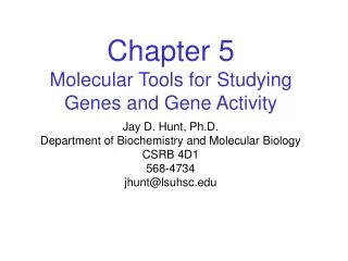
Chapter 16: The Molecular Basis of Inheritance. Chapter 14 – traits carried in gametes Chapter 15 – genes found on chromosomes Chapter 16 – DNA is the molecule of inheritance How did we learn this?.

E N D
Chapter 16: The Molecular Basis of Inheritance Chapter 14 – traits carried in gametes Chapter 15 – genes found on chromosomes Chapter 16 – DNA is the molecule of inheritance How did we learn this?
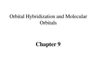
Bacteria of the “S” (smooth) strain of Streptococcus pneumoniae are pathogenic because they have a capsule that protects them from an animal’s defense system. Bacteria of the “R” (rough) strain lack a capsule and are nonpathogenic. Frederick Griffith injected mice with the two strains as shown below: CONCLUSION EXPERIMENT RESULTS Living S (control) cells Living R (control) cells Heat-killed (control) S cells Mixture of heat-killed S cells and living R cells Mouse dies Mouse healthy Mouse healthy Mouse dies Living S cells are found in blood sample. Griffith concluded that the living R bacteria had been transformed into pathogenic S bacteria by an unknown, heritable substance from the dead S cells. Chapter 16: The Molecular Basis of Inheritance • What evidence did Frederick Griffith have that DNA was the genetic material? Transformation – external DNA assimilated by a cell which changes the cell’s genotype and phenotype
Phage head Tail Tail fiber DNA 100 nm Bacterial cell Chapter 16: The Molecular Basis of Inheritance • What evidence did Frederick Griffith have that DNA was the genetic material? • How did Hershey & Chase determine that DNA was the genetic material? • - Bacteriophage – viruses that infect bacteria
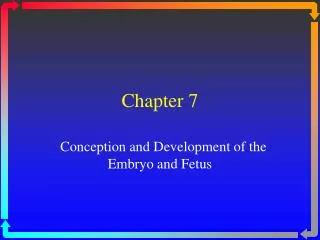
EXPERIMENT In their famous 1952 experiment, Alfred Hershey and Martha Chase used radioactive sulfur and phosphorus to trace the fates of the protein and DNA, respectively, of T2 phages that infected bacterial cells. 3 2 Agitated in a blender to separate phages outside the bacteria from the bacterial cells. Centrifuged the mixture so that bacteria formed a pellet at the bottom of the test tube. 4 1 Measured the radioactivity in the pellet and the liquid Mixed radioactively labeled phages with bacteria. The phages infected the bacterial cells. Radioactive protein Empty protein shell Radioactivity (phage protein) in liquid Phage Bacterial cell DNA Batch 1: Phages were grown with radioactive sulfur (35S), which was incorporated into phage protein (pink). Phage DNA Centrifuge Radioactive DNA Pellet (bacterial cells and contents) Batch 2: Phages were grown with radioactive phosphorus (32P), which was incorporated into phage DNA (blue). Centrifuge Radioactivity (phage DNA) in pellet Pellet Chapter 16: The Molecular Basis of Inheritance • What evidence did Frederick Griffith have that DNA was the genetic material? • How did Hershey & Chase determine that DNA was the genetic material?
CONCLUSION Phage proteins remained outside the bacterial cells during infection, while phage DNA entered the cells. When cultured, bacterial cells with radioactive phage DNA released new phages with some radioactive phosphorus. Hershey and Chase concluded that DNA, not protein, functions as the T2 phage’s genetic material. RESULTS Chapter 16: The Molecular Basis of Inheritance • What evidence did Frederick Griffith have that DNA was the genetic material? • How did Hershey & Chase determine that DNA was the genetic material?
Sugar-phosphate backbone Nitrogenous bases 5 end CH3 O– 5 O H CH2 O P O O 1 4 N O– N H H H H H O 2 3 H Thymine (T) O H H CH2 O O P N O N H O– H N H H H N N H H Adenine (A) H H O H N CH2 O O P H O O– N H N H H H O H Cytosine (C) O 5 H CH2 O N P O O O 1 4 O– H N H Phosphate H H N 2 H 3 DNA nucleotide N H OH N Sugar (deoxyribose) 3 end H H Guanine (G) Chapter 16: The Molecular Basis of Inheritance • What evidence did Frederick Griffith have that DNA was the genetic material? • How did Hershey & Chase determine that DNA was the genetic material? • What was Chargaff’s contribution? • Nucleotide composition • Human • A – 30.3% • T – 30.3% • C – 19.9% • G – 19.5% Chargaff’s rules A=T C=G Note: 5’ end – phosphate group 3’ end - -OH
Franklin’s X-ray diffraction Photograph of DNA (b) (a) Rosalind Franklin Chapter 16: The Molecular Basis of Inheritance • What evidence did Frederick Griffith have that DNA was the genetic material? • How did Hershey & Chase determine that DNA was the genetic material? • What was Chargaff’s contribution? • What experiment did Watson & Crick do? • - NONE – compiled everyone else’s data Double helix
Purine + Purine: too wide Pyrimidine + pyrimidine: too narrow Purine + pyrimidine: width Consistent with X-ray data Chapter 16: The Molecular Basis of Inheritance • What evidence did Frederick Griffith have that DNA was the genetic material? • How did Hershey & Chase determine that DNA was the genetic material? • What was Chargaff’s contribution? • What experiment did Watson & Crick do? • NONE – compiled everyone else’s data • Determined double helix from Rosalind Franklin’s data
N O H CH3 N N N N H Sugar N N O Sugar Adenine (A) Thymine (T) H O N H N N N H N Sugar N N N O H Sugar H Cytosine (C) Guanine (G) Chapter 16: The Molecular Basis of Inheritance • What evidence did Frederick Griffith have that DNA was the genetic material? • How did Hershey & Chase determine that DNA was the genetic material? • What was Chargaff’s contribution? • What experiment did Watson & Crick do? • NONE – compiled everyone else’s data • Determined double helix from Rosalind Franklin’s data
5 end G C O OH A T Hydrogen bond P 3 end –O O T A OH O H2C A T 1 nm O O CH2 O C G P O O– –O 3.4 nm O P C G O O H2C O G C A T O CH2 O O G C P O O– –O O P O O H2C O G C T A O CH2 O O P T A O –O O– O P O A T O H2C O A T T A O CH2 OH O O– 3 end P O G C O 0.34 nm T 5 end A (a) Key features of DNA structure (b) Partial chemical structure (c) Space-filling model Figure 16.7 The double helix DNA is antiparallel Hydrogen bonds hold double helix together Now that we know how DNA is shaped & how it fits together…..
Announcements • Test corrections • Stapled behind • Place in box • Canned food – no later than Thursday • Pie Festival – for Ch 16 – 18 test (instead of cans)
T A A A A T A T T T T A G C G C C C C G G G G C A A T T T T A A A A T T T A A A A T A T T T T A C G G G G C G C C C C G (a) The parent molecule has two complementary strands of DNA. Each base is paired by hydrogen bonding with its specific partner, A with T and G with C. (c) Each parental strand now serves as a template that determines the order of nucleotides along a new, complementary strand. (d) The nucleotides are connected to form the sugar-phosphate backbones of the new strands. Each “daughter” DNA molecule consists of one parental strand and one new strand. (b) The first step in replication is separation of the two DNA strands. Chapter 16: The Molecular Basis of Inheritance • What evidence did Frederick Griffith have that DNA was the genetic material? • How did Hershey & Chase determine that DNA was the genetic material? • What was Chargaff’s contribution? • What experiment did Watson & Crick do? • How does DNA replicate itself? The basic concept But is replication done in a conservative, semi-conservative or dispersive manner?
First replication Second replication Parent cell (a) Conservative model. The two parental strands reassociate after acting as templates for new strands, thus restoring the parental double helix. (b) Semiconserva- tive model. The two strands of the parental molecule separate, and each functions as a template for synthesis of a new, comple- mentary strand. (c) Dispersive model. Each strand of both daughter mol- ecules contains a mixture of old and newly synthesized DNA. Figure 16.10 Three alternative models of DNA replication • Conservative • Original strands • return together • Semi-conservative • One original & • one new strand • Dispersive • Original strands • are dispersed • through new • strands The Meselson – Stahl Experiment showed how DNA replicates.
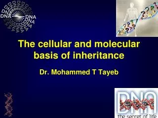
EXPERIMENT Matthew Meselson and Franklin Stahl cultured E. coli bacteria for several generations on a medium containing nucleotide precursors labeled with a heavy isotope of nitrogen, 15N. The bacteria incorporated the heavy nitrogen into their DNA. The scientists then transferred the bacteria to a medium with only 14N, the lighter, more common isotope of nitrogen. Any new DNA that the bacteria synthesized would be lighter than the parental DNA made in the 15N medium. Meselson and Stahl could distinguish DNA of different densities by centrifuging DNA extracted from the bacteria. Bacteria cultured in medium containing 15N Bacteria transferred to medium containing 14N 2 1 3 4 RESULTS Less dense DNA sample centrifuged after 40 min (after second replication) DNA sample centrifuged after 20 min (after first replication) More dense The bands in these two centrifuge tubes represent the results of centrifuging two DNA samples from the flask in step 2, one sample taken after 20 minutes and one after 40 minutes. Figure 16.11 Does DNA replication follow the conservative, semiconservative, or dispersive model?
CONCLUSION Meselson and Stahl concluded that DNA replication follows the semiconservative model by comparing their result to the results predicted by each of the three models in Figure 16.10. The first replication in the 14N medium produced a band of hybrid (15N–14N) DNA. This result eliminated the conservative model. A second replication produced both light and hybrid DNA, a result that eliminated the dispersive model and supported the semiconservative model. First replication Second replication Conservative model Semiconservative model Dispersive model
Chapter 16: The Molecular Basis of Inheritance • What evidence did Frederick Griffith have that DNA was the genetic material? • How did Hershey & Chase determine that DNA was the genetic material? • What was Chargaff’s contribution? • What experiment did Watson & Crick do? • How does DNA replicate itself? • How is DNA replicated? • Origin of replication - sequence of DNA
Chapter 16: The Molecular Basis of Inheritance • What evidence did Frederick Griffith have that DNA was the genetic material? • How did Hershey & Chase determine that DNA was the genetic material? • What was Chargaff’s contribution? • What experiment did Watson & Crick do? • How does DNA replicate itself? • How is DNA replicated? • Origin • Elongation • DNA polymerase • 500 bp/sec bacteria, 50 bp/sec in us • New base added to 3’end (-OH) • dNTPs provide energy
New strand Template strand 3’ end 5’ end 3’ end 5’ end Sugar A T A T Base Phosphate C G C G G G C C A T A T OH P P P P 3’ end P Pyrophosphate C C OH 2 P Nucleoside triphosphate 5’ end 5’ end Figure 16.13 Incorporation of a nucleotide into a DNA strand OH DNA polymerase
Chapter 16: The Molecular Basis of Inheritance • What evidence did Frederick Griffith have that DNA was the genetic material? • How did Hershey & Chase determine that DNA was the genetic material? • What was Chargaff’s contribution? • What experiment did Watson & Crick do? • How does DNA replicate itself? • How is DNA replicated? • Origin • Elongation • Dealing with antiparallel strands
DNA pol Ill elongates DNA strands only in the 5 3 direction. One new strand, the leading strand, can elongate continuously 5 3 as the replication fork progresses. 2 3 1 4 3 5 Parental DNA 5 The other new strand, the lagging strand must grow in an overall 3 5 direction by addition of short segments, Okazaki fragments, that grow 5 3 (numbered here in the order they were made). 3 Okazaki fragments 2 3 1 5 DNA pol III Template strand DNA ligase joins Okazaki fragments by forming a bond between their free ends. This results in a continuous strand. Leading strand Lagging strand 3 1 2 Template strand DNA ligase Overall direction of replication Figure 16.14 Synthesis of leading and lagging strands during DNA replication
6 7 5 1 2 3 4 3 5 3 5 Templatestrand DNA pol III adds DNA nucleotides to the primer, forming an Okazaki fragment. Primase joins RNA nucleotides into a primer. RNA primer 3 5 3 1 5 After reaching the next RNA primer (not shown), DNA pol III falls off. Okazakifragment 3 3 5 1 5 After the second fragment is primed. DNA pol III adds DNAnucleotides until it reaches the first primer and falls off. 5 3 3 2 5 1 DNA pol 1 replaces the RNA with DNA, adding to the 3 end of fragment 2. 5 3 3 5 2 1 DNA ligase forms a bond between the newest DNAand the adjacent DNA of fragment 1. The lagging strand in this region is nowcomplete. 5 3 3 2 5 1 Overall direction of replication Figure 16.15 Synthesis of the lagging strand
Announcements • Learning log – available – greater percentage of “critical thinking” questions • People are hungry!!!!! – tardies????
Overall direction of replication Lagging strand Leading strand Origin of replication Helicase unwinds the parental double helix. 1 2 Molecules of single- strand binding protein stabilize the unwound template strands. The leading strand is synthesized continuously in the 5 3 direction by DNA pol III. 3 Leading strand Lagging strand OVERVIEW DNA pol III Leading strand 5 Replication fork DNA ligase DNA pol I 3 Primase 2 Parental DNA DNA pol III Lagging strand 1 Primer 3 4 Primase begins synthesis of RNA primer for fifth Okazaki fragment. 3 5 4 5 6 7 DNA pol III is completing synthesis of the fourth fragment, when it reaches the RNA primer on the third fragment, it will dissociate, move to the replication fork, and add DNA nucleotides to the 3 endof the fifth fragment primer. DNA ligase bonds the 3 end of the second fragment to the 5 end of the first fragment. DNA pol I removes the primer from the 5 end of the second fragment, replacing it with DNA nucleotides that it adds one by one to the 3’ end of the third fragment. The replacement of the last RNA nucleotide with DNA leaves the sugar- phosphate backbone with a free 3 end. Figure 16.16 A summary of bacterial DNA replication
Chapter 16: The Molecular Basis of Inheritance • What evidence did Frederick Griffith have that DNA was the genetic material? • How did Hershey & Chase determine that DNA was the genetic material? • What was Chargaff’s contribution? • What experiment did Watson & Crick do? • How does DNA replicate itself? • How is DNA replicated? • How is DNA repaired? • Proofreading – mismatch repair – DNA polymerase fixes mistakes made • during copying • Excision repair
A thymine dimer distorts the DNA molecule. 1 3 4 2 A nuclease enzyme cuts the damaged DNA strand at two points and the damaged section is removed. Nuclease Repair synthesis by a DNA polymerase fills in the missing nucleotides. DNA polymerase DNA ligase DNA ligase seals the Free end of the new DNA To the old DNA, making the strand complete. Figure 16.17 Nucleotide excision repair of DNA damage
Chapter 16: The Molecular Basis of Inheritance • What evidence did Frederick Griffith have that DNA was the genetic material? • How did Hershey & Chase determine that DNA was the genetic material? • What was Chargaff’s contribution? • What experiment did Watson & Crick do? • How does DNA replicate itself? • How is DNA replicated? • How is DNA repaired? • What happens at the end of chromosomes (telomere)? • Recall DNA polymerase works 5’ → 3’ direction • After RNA primer is removed, chromosome is shortened
5 End of parental DNA strands Leading strand Lagging strand 3 Last fragment Previous fragment RNA primer 5 Lagging strand 3 Primer removed but cannot be replaced with DNA because no 3 end available for DNA polymerase Removal of primers and replacement with DNA where a 3 end is available 5 3 Second round of replication 5 New leading strand 3 New lagging strand 5 3 Further rounds of replication Shorter and shorter daughter molecules Figure 16.18 Shortening of the ends of linear DNA molecules
Telomerase • active in germ line cells & newborns • to prevent shortening • -extends telomere a bit • -has its own RNA • - complementary to telomere • - serves as primer 2009 Nobel Prize in Medicine - for the discovery of how chromosomes are protected by telomeres and the enzyme telomerase