Download
1 / 232
2.41k likes | 2.78k Views
CARCINOGENESIS: THE MOLECULAR BASIS OF CANCER. Nonlethal genetic damage lies at the heart of carcinogenesis. Mutation) may be acquired by the action of environmental agents, such as chemicals, radiation, or viruses, or it may be inherited in the germ line.

E N D
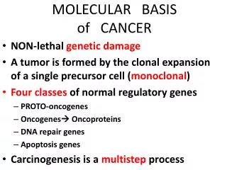
Nonlethal genetic damage lies at the heart of carcinogenesis. • Mutation) may be acquired by the action of environmental agents, such as chemicals, radiation, or viruses, or it may be inherited in the germ line.
The genetic hypothesis of cancer implies that a tumor mass results from the clonal expansion of a single progenitor cell that has incurred genetic damage (i.e., tumors are monoclonal). • Clonality of tumors is assessed readily in women who are heterozygous for polymorphic X-linked markers, such as the enzyme glucose-6-phosphate dehydrogenase or X-linked restriction-fragment-length polymorphisms.
Four classes of normal regulatory genes are involved : • 1-growth-promoting proto-oncogenes, • 2-growth-inhibiting tumorsuppressor genes, • 3-genes that regulate apoptosis • 4-genes involved in DNA
Mutant alleles of proto-oncogenes are called oncogenes. • They are considered dominant because mutation of a single allele can lead to cellular transformation. • Both normal alleles of tumor suppressor genes must be damaged for transformation to occur, referred to as recessive oncogenes.
Genes that regulate apoptosis may be dominant, as are proto-oncogenes, or they may behave as tumor suppressor genes (recessive ).
Tumor suppressor genes are of 2 types : • 1- promoters genes • 2- caretakers genes
Promoters are the traditional tumor suppressor genes, such as RB or p53, • mutation of these genes leads to cell transformation by releasing the control on cellular proliferation.
Caretaker genes are responsible for processes that ensure the integrity of the genome, such as DNA repair. • Mutation of caretaker genes does not directly transform cells by affecting proliferation or apoptosis. • DNA repair genes affect cell proliferation or survival indirectly by influencing the ability to repair nonlethal damage in other genes, including proto-oncogenes, tumor suppressor genes, and genes that regulate apoptosis.
Carcinogenesis is a multistep process at both the phenotypic and the genetic levels, resulting from the accumulation of multiple mutations. • Malignant neoplasms have several phenotypic attributes, such as excessive growth, local invasiveness, and the ability to form distant metastases.
Tumor progression over a period of time, many tumors become more aggressive and acquire greater malignant potential which is not simply represented by an increase in tumor size.
Tumor progression and associated heterogeneity results from multiple mutations that accumulate independently in different tumor cells, generating subclones with different characteristics
Even though most malignant tumors are monoclonal in origin, by the time they become clinically evident, their constituent cells are extremely heterogeneous. • During progression, tumor cells are subjected to immune and nonimmune selection pressures. • E.g cells that are highly antigenic are destroyed by host defenses, whereas those with reduced growth factor requirements are positively selected. • A growing tumor tends to be enriched for subclones that are capable of survival, growth, invasion, and metastasis.
Features of malignent cells • 1-Self-sufficiency in growth signals • 2-Insensitivity to growth-inhibitory signals • 3-Evasion of apoptosis • 4-Limitless replicative potential (i.e., overcoming cellular senescence and avoiding mitotic catastrophe) • 5-Development of sustained angiogenesis • 6-Ability to invade and metastasize • 7-Genomic instability resulting from defects in DNA repair
Self-Sufficiency in Growth Signals • Genes that promote autonomous cell growth in cancer cells are called oncogenes. • They are derived by mutations in proto-oncogenes and are characterized by the ability to promote cell growth in the absence of normal growth-promoting signals. • Their products, called oncoproteins, resemble the normal products of proto-oncogenes except that oncoproteins are devoid of important regulatory elements, and their production in the transformed cells does not depend on growth factors or other external signals.
The binding of a growth factor to its specific receptor on the cell membrane causes transient and limited activation of the growth factor receptor. • activates several signal-transducing proteins on the inner leaflet of the plasma membrane • transmission of the transduced signal across the cytosol to the nucleus via second messengers or a cascade of signal transduction molecules • induction and activation of nuclear regulatory factors that initiate DNA transcription • progression of the cell into the cell cycle, resulting ultimately in cell division
Growth Factors • All normal cells require stimulation by growth factors to undergo proliferation. • Types : • 1- paracrine action. growth factors are made by one cell type and act on a neighboring cell to stimulate proliferation 2-autocrine action Many cancer cells acquire growth self-sufficiency by acquiring the ability to synthesize the same growth factors to which they are responsive.
Glioblastomas secrete platelet-derived growth factor (PDGF) and express the PDGF receptor, • Many sarcomas make both transforming growth factor-α (TGF-α) and its receptor. • Genes that encode homologues of fibroblast growth factors (e.g., hst-1 and FGF3) have been detected in several gastrointestinal and breast tumors; • FGF-2 is expressed in human melanomas but not normal melanocytes.
Hepatocyte growth factor (HGF) and its receptor c-Met are both overexpressed in follicular carcinomas of the thyroid. • In many instances the growth factor gene itself is not altered or mutated, but the products of other oncogenes (e.g., RAS) stimulate overexpression of growth factor genes and the subsequent development of an autocrine loop.
Growth Factor Receptors • Mutant receptor proteins deliver continuous mitogenic signals to cells, even in the absence of the growth factor in the environment. • overexpression of growth factor receptors can render cancer cells hyper-responsive to levels of the growth factor that would not normally trigger proliferation.
E.g • overexpression involve the epidermal growth factor (EGF) receptor family. ERBB1, • the EGF receptor, is overexpressed in 80% of squamous cell carcinomas of the lung. • In 50% or more of glioblastomas. • In 80-100% of epithelial tumors of the head and neck.
HER2/NEU (ERBB2), is amplified in 25-30% of breast cancers and adenocarcinomas of the lung, ovary, and salivary glands. • These tumors are exquisitely sensitive to the mitogenic effects of small amounts of growth factors • High level of HER2/NEU protein in breast cancer cells is a poor prognosis.
The significance of HER2/NEU in the pathogenesis of breast cancers is illustrated by the clinical benefit derived from blocking the extracellular domain of this receptor with anti-HER2/NEU antibodies. • Treatment of breast cancer with anti-HER2/NEU antibody (herciptin ) proved to be clinically effective .
Signal-Transducing Proteins • These signaling molecules couple growth factor receptors to their nuclear targets. • Many such signaling proteins are associated with the inner leaflet of the plasma membrane, where they receive signals from activated growth factor receptors and transmit them to the nucleus, either through second messengers or through a cascade of phosphorylation and activation of signal transduction molecules. • Two important members in this category are • 1-RAS gene • 2-ABL gene
RAS is the most commonly mutated proto-oncogene in human tumors. • Approximately 30% of all human tumors contain mutated versions of the RAS gene • The incidence is even higher in some specific cancers (e.g., colon and pancreatic adenocarcinomas). • RAS is a member of a family of small G proteins that bind guanosine nucleotides (guanosine triphosphate [GTP] and guanosine diphosphate [GDP]).
Normal RAS proteins flip back and forth between an excited signal-transmitting state and a quiescent state. • RAS proteins are inactive when bound to GDP • stimulation of cells by growth factors leads to exchange of GDP for GTP and subsequent activation of RAS.
The activated RAS in turn stimulates down-stream regulators of proliferation, such as the RAF-mitogen-activated protein (MAP) kinase mitogenic cascade, which floods the nucleus with signals for cell proliferation. • The excited signal-emitting stage of the normal RAS protein is short-lived • Intrinsic guanosine triphosphatase (GTPase) activity hydrolyzes GTP to GDP, releasing a phosphate group and returning the protein to its quiescent inactive state.
The GTPase activity of activated RAS protein is magnified dramatically by a family of GTPase-activating proteins (GAPs), which act as molecular brakes that prevent uncontrolled RAS activation by favoring hydrolysis of GTP to GDP.
The RAS gene is most commonly activated by point mutations. • Point mutations can affect : • 1-GTP-binding pocket • 2-the enzymatic region essential for GTP hydrolysis. • Mutations at these locations interfere with GTP hydrolysis that is essential to convert RAS into an inactive form. • RAS is thus trapped in its activated GTP-bound form, and the cell is forced into a continuously proliferating state.
mutations in RAS protein would be mimicked by mutations in the GAPs that fail to restrain normal RAS proteins. • E.g mutation of neurofibromin 1, a GAP, is associated with familial neurofibromatosis type 1
The ABL proto-oncogene has tyrosine kinase activity that is dampened by internal negative regulatory domains. • In chronic myeloid leukemia (CML) and acute lymphocytic leukemias, • When ABL gene is translocated from its normal site on chromosome 9 to chromosome 22, where it fuses with part of the breakpoint cluster region (BCR) gene = Philadelphia (Ph) chromosome .
The BCR-ABL hybrid protein has potent, unregulated tyrosine kinase activity, which activates several pathways, including the RAS-RAF cascade. • Normal ABL protein localizes in the nucleus, where its role is to promote apoptosis of cells that suffer DNA damage. • The BCR-ABL gene cannot perform this function, because it is retained in the cytoplasm as a result of abnormal tyrosine kinase activity.
A cell with BCR-ABL fusion gene is dysregulated in two ways: • 1-inappropriate tyrosine kinase activity leads to growth autonomy. • 2- impairment of apoptosis.
The crucial role of BCR-ABL in transformation has been confirmed by the dramatic clinical response of patients with chronic myeloid leukemia after therapy with an inhibitor of the BCR-ABL fusion kinase called imatinib mesylate (Gleevec).
Nuclear Transcription Factors • Growth autonomy may occur as a consequence of mutations affecting genes that regulate transcription of DNA. • MYC, MYB, JUN, FOS, and REL oncogenes, function as transcription factors that regulate the expression of growth-promoting genes, such as cyclins.
the MYC gene is involved most commonly in human tumors. • The MYC proto-oncogene is expressed in virtually all cells • the MYC protein is induced rapidly when quiescent cells receive a signal to divide.
In normal cells, MYC levels decline to near basal level when the cell cycle begins. • In contrast, oncogenic versions of the MYC gene are associated with persistent expression or overexpression, contributing to sustained proliferation.
The MYC protein can either activate or repress the transcription of other genes. • Those activated by MYC include several growth-promoting genes, including cyclin-dependent kinases (CDKs), whose products drive cells into the cell cycle. • Genes repressed by MYC include the CDK inhibitors (CDKIs).
MYC promotes tumorigenesis by increasing expression of genes that promote progression through the cell cycle and repressing genes that slow or prevent progression through the cell cycle.
Dysregulation of the MYC gene resulting from a t(8;14) translocation occurs in Burkitt lymphoma, a B-cell tumor. • MYC is also amplified in breast, colon, lung, and many other cancers; • N-MYC and L-MYC genes are amplified in neuroblastomas and small-cell cancers of lung.
Cyclins and Cyclin-Dependent Kinases (CDKs) • Cancers may become autonomous if the genes that drive the cell cycle become dysregulated by mutations or amplification. • Progression of cells through the various phases of the cell cycle is controlled by CDKs. • CDKs are activated by binding to cyclins, so called because of the cyclic nature of their production and degradation.
The CDK-cyclin complexes phosphorylate crucial target proteins that drive the cell through the cell cycle. • On completion of this task, cyclin levels decline rapidly. • More than 15 cyclins have been identified; cyclins D, E, A, and B appear sequentially during the cell cycle and bind to one or more CDK.
Mishaps affecting the expression of cyclin D or CDK4 seem to be a common event in neoplastic transformation. • The cyclin D genes are overexpressed in many cancers, including those affecting the breast, esophagus, liver, and a subset of lymphomas.