Download

1 / 52
580 likes | 903 Views
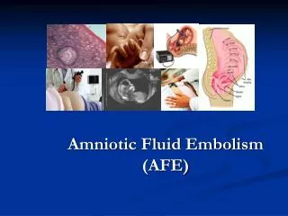
This comprehensive overview details the human placenta's development, structure, and pivotal functions, including gas and nutrient exchange between mother and fetus. The placenta is formed from fetal and maternal components and has distinct anatomical features, such as its discoid shape and two surfaces. Critical insights into placental abnormalities, the role of amniotic fluid, and the mechanisms governing the transfer of substances are also discussed. This resource is essential for understanding placental physiology and its impact on fetal development.

E N D
Placenta and Amniotic fluid- Structure, Function, and Abnormalities
Placenta • Human placenta develops from two sources Fetal component- Chorionic frondosum Maternal component- decidua basalis • Placental development begins at 6 weeks and is completed by 12 th week
Human placenta is • Discoid in shape • Haemochorial • Deciduate
Placenta at Term- Gross Anatomy • Fleshy • Weight-500gm • Diameter- 15-20 cm • Thickness-2.5 cm • Spongy to feel • Occupies 30% of the uterine wall • Two surfaces- Maternal and fetal • 4/5th of the placenta is of fetal origin and 1/5 is of maternal origin
Fetal surface of the placenta • Covered by smooth and glistening amnion overlying the chorion • Umbilical cord is attached at or near its centre • Branches of the umbilical vessels are visible beneath the amnion as they radiate from the insertion of the cord
Maternal surface of the placenta • Rough and spongy • Maternal blood gives it dull red colour • Remanants of the decidua basalis gives it shaggy appearance • Divided into 15-20 cotyledons by the septa
Margins of the placenta are formed by fused chorionic and the basal plate • Placenta is attached to the upper part of the uterine body either at the posterior or anterior wall • After delivery ,placenta separates with the line of separation being through decidua spongiosum (intermediate spongy layer of the decidua basalis
Structure of the placenta • Placenta is limited by the amniotic membrane on the fetal side and by the basal plate on the maternal • Between these two lies the intervillous space filled with maternal blood and stem villi with their branches
Amniotic membrane- single layer of cubical epithelium loosely attached to adjacent chorionic plate and does not take part in placental formation • Chorionic plate- forms the roof of the placenta • From outside inwards consists of • Syncitotrophoblast • Cytotrophoblast • Extraembryonic mesoderm with branches of umbilical vessels
Basal Plate- forms the floor From outside inwards it consist of • Compact and spongy layer of decidua basalis • Layer of Nitabuch • Cytotrophoblastic shell • Syncytiotrophoblast Basal plate is perforated by the spiral arteries allowing entry of maternal blood into intervillous space
Layer of Nitabuch - is a fibrinous layer formed at the junction of cytotrohoblastic shell with decidua due to fibrinoid degeneration of syncitotrohoblast • It prevents excessive penetration of the decidua by the trophoblast • Nitabuch membrane is absent in placenta accreta and other morbidly adherent placentas
Intervillous space: • Numerous branch villi arising from the stem villi project into this space • It is lined internally on all sides by the syncytiotrophoblast and is filled with maternal blood
Stem (Anchoring villi ) • Arise from the chorionic plate and extend to the basal plate • Fetal cotyledon (60-100 ) – derived from one major primary stem villus and is the structural unit of placenta • Maternal cotyledon (15-20 ) contains 3-5 fetal cotyledons • Villus is the functional unit of placenta • Total surface of the villi for exchange varies between 4-14 sq meters
Placental barrier or membrane Maternal and fetal blood are separated by placental membrane or barrier (0.025 mm thick ) • Endothelial lining of fetal vessels • Connective tissue of the villi • Basement membrane • Cytotrophoblast • Syncytiotrophoblast
Placental Function • Transfer of gases ,nutrients and waste products , namely • Respiratory function • Nutritive function • Excretory function • Endocrine and enzymatic function • Barrier function • Immulogical function
Factors affecting the transfer between mother and the fetus • Physical properties of the substance- molecular weight, lipid solubility, ionised substances • Area and functional integrity of the placental membrane • Rate of blood flow • Concentration gradient of the substance on either side of the exchange membrane
Mechanism involved in the transfer of substances • Simple diffusion-O2 and CO2 • Facilitated diffusion ( carrier mediated ) –glucose ,vitamins • Active transfer ( against concentration gradient )-ions • Endocytosis- invagination of cell membrane to form intracellular vesicle • Endocytosis-Release of substances in the vesicles to extracellular space eg IgG immunoglobulin
Respiratory function • Although fetal respiratory movement occurs, no active exchange of gases takes place • Intake of oxygen and output of carbon dioxide take place by simple diffusion across the fetal membrane • O2 delivery to the fetus is at the rate of 8 ml/kg which is achieved by cord blood flow of 160-320ml/min
Excretory function • Waste products from the fetus such as urea, uric acid, cretinine are excreted to the maternal blood by simple diffusion
Nutritive function Fetus obtains its nutrients from the maternal blood • Glucose- transferred to the fetus by facilitated diffusion • Lipids for fetal growth and development has dual origin. They are transferred across the fetal membrane or synthesised in the fetus • Amino acids are transferred by active transport • Water and electrolytes- Na, K ,Cl cross by simple diffusion, Ca , P, and Fe cross by active transport • Water soluble vitamins are transferred by active transport but the fat soluble vitamins are transferred slowly
Barrier Function • Placental membrane is thought to be a protective barrier for the fetus against harmful agents in the maternal blood • Substances with large molecular weight or size like insulin or heparin are transferred minimally • Only IgG ( not IgA or Ig M )antibodies and antigens can cross the placental barrier • Most drugs can cross the placental barrier and some can be teratogenic • Various viruses, bacteria, protozoa can cross the placenta and affect the fetus in utero
Immunological function • Inspite of foreign paternally inherited antigens in the fetus and placenta, there is no graft rejection due to immunological protection provided by the placenta
Endocrine and Enzymatic function • Placenta secretes various hormones – Protein hormones like HCG, human placental lactogen,pregnancy specific beta 1 glycoprotein,,pregnancy associated plasma protein, steroidal hormones like estrogen and progestrone • Enzymes secreted are diamine oxidase-which activates the circulatory pressor amines,oxytocinase which neutralizes oxytocin, phospholipase A2 which synthesizes arachidonic acid
Placental abnormalities Placenta succenturiata (3%) • One or more small lobe or cotyledon of placenta may be placed at a varying distance from the main placental margin • A leash of vessels connecting the main to the small lobe traverse through the membranes • Accessory lobe is developed from activated villi on the chorionic laeve
Clinical significance- If succenturiate lobe is retained following birth of placenta it may lead to • PPH • Subinvolution • Uterine sepsis • Poly formation Treatment- exploration of the uterus and removal of the lobe
Circumvallate placenta Development- • Due to smaller chorionic plate than the basal plate • The chorionic plate does not extend into the placenta margin • The amnion and chorion are folded and rolled back to form a ring leaving a rim of uncovered placental tissue
Morphology • Fetal surface has a central depressed zone surrounded by a usually complete thickened white ring made up of double fold of amnion and chorion • Branching vessels radiate from the cord insertion upto ring only • Area outside the ring is thicker, elevated and rounded
Clinical significance • There are more chances of – • Miscarriage • Hydrorrhoea gravidarum • Antepartum haemorrhage • Preterm delivery • Fetal growth restriction • Retained placenta or membrane
Placenta marginata • A thin fibrous ring is present at the margin of the chorionic plate where the fetal vessels appear to terminate
Membranous placenta • The whole of the chorion is covered by functioning villi and thus placenta appears as thin membranous structure on ultrasonography
Chorioangioma • Are the most common benign tumors of the placenta and are hamartomas of primitive chorionic mesenchyme • Small tumors may be asymptomatic but large tumors may be associated with hydroamnios and antepartum haemorrhage
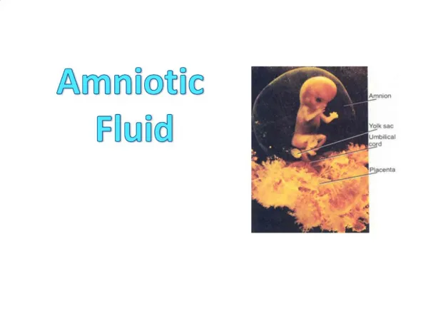
Amniotic fluid • It is the fluid in the amniotic sac surrounding the fetus • Origin – both mother and fetus • Transudation from maternal circulation across the placental surface and fetal membranes • Active secretion from amniotic epithelium • Transudation across surface of umbilical cord and fetal placental circulation • Contribution from fetal urine • Tracheobronchial secretion • Transfer across fetal skin
Volume- varies according to the gestational age • Measures • 12 weeks – 50 ml • 20 weeks- 400 ml • 36 weeks- 800ml-1 liter • At term - it reduces to apprx 700ml
Composition- Organic constituents • Proteins-0.3 mg/dl • Glucose- 20mg/dl • Urea- 30 mg/dl • Non protein nitrogen-30mg/dl • Uric acid – 4 mg/dl • Creatinine -2 mg/dl • Lipids- 50 mg/ dl • Hormones- insulin,prolactin, renin Inorganic constituents- Na, K,Cl Suspended particles- Lanugo,Desqamated fetal skin cells,vernix caseosa,shedded amniotic cells, cells from thr respiratory tract,GIT,Genitourinary tract
Physical features • Faintly alkaline • Low specific gravity-1.010 • Becomes highly hypotonic to maternal serum at term pregnancy • Osmolarity of 250 mOsmol/liter is suggestive of fetal maturity
Colour • In early pregnancy it is colourless • At term becomes pale straw coloured due to preence of exfoliated lanugo and epidermal cells from fetal skin
Abnormal appearance • Greenish- due to presence of meconium • Golden yellow-due to presence of bilirubin resulting from fetal cell hemolysis due to Rh incompatibility • Greenish yellow- in post maturity • Dark maroon/ blood stained – due to altered blood in accidental haemorrhage • Prune juice/dark brown- in presence of retained dead fetus
Functions of amniotic fluid During pregnancy • Act as a shock absorber to protect the fetus from external injury • Maintains the fetal temprature • Allows free movement and growth of fetus • Prevents adhesion formation between the fetal parts and the amniotic sac • Has some nutritive value because of small amount of protein and salt content
During Labour • It forms hydrostatic wedge to help dilatation of cervix • During uterine contractions , the amniotic fluid in the intact membranes prevents interference with placental circulation • Provides pool for the fetus to excrete urine • Protect the fetus from the ascending infections by its bactercidal action
Clinical importance • Study of amniotic fluid helps in knowing the well being and maturity of fetus • Intramniotic instillation of prostaglandins and hypertonic saline can be used for induction of abortion • Artificial rupture of membranes to drain liquor is a method of induction and augmentation of labour • Excess liquor (polyhydroamnios), less liquor known as (oligohydroamnios ) can be estimated by ultrasound measurement of amniotic fluid index (AFI )
Measurment of AF • Measurement of AFI- quantitative method of measurement of amniotic fluid by usg. Single largest pocket is measured in four quadrants and added. • Normal range is 5-24 cm • Single deepest pocket • Normal range is 2-8 cm
Polyhydroamnios • Defined as excess of amniotic fluid of more than 2000ml or AFI> 25 cm or SDP>8cm
Etiology • Idiopathic- seen in 2/3rd of the cases Fetal causes- Anencephaly spina bifida Esophageal and duodenal atresia Facial cleft and neck masses Congenital diaphragmatic hernia Fetal sacrococcygeal teratoma Fetal infections Hydrops fetalis Multiple pregnancy
Placental causes- choriangioma of the placenta • Maternal causes- Diabetes, cardiac or renal disease
Types • Acute- sudden increase • Chronic- gradual increase
Symptoms- breathlessness due to mechnacial compression, edema of legs, varicosities in legs, • Signs-Abdomen is markedly distended, skin is tense,shiny fundal height >POG,
Complications Maternal During pregnancy- • Incresed incidence of preeclampsia • Malpresentation • Premature rupture of membranes • Preterm labour • Abruptio placentae • Cardiorespiratory embrassment
During labour Premature rupture of membranes Cord prolapse Uterine inertia PPH Puerperium Subinvolution Puerperal sepsis Fetal Complications • High perinatal mortality due to prematurity and congenital malformations
Management • Rule out fetal congenital anomalies • Bed rest • Amnioreduction- 1-1.5 liters of amniotic fluid is removed over 3 hours to relieve maternal distress • Indomethacin therapy- impairs lung fluid production,enhances absorption of amniotic fluid, decreases fetal urine production,increases fluid movement across fetal membranes • Dose – 1.5-3 mg/kg from 24-35 weeks for 2 weeks • S/E- premature closure of patent ductus arterious
Oligohydroamnios • Amniotic fluid is less than 200 ml at term or AFI < 5 cm OR SDP< 2 cm Etiology • Fetal chromosomal anomalies • Intrauterine infections • Drugs- PG inhibitors, ACE inhibitors • Renal agenesis or obstruction of the urinary tract • IUGR associated with placental insufficency • Amnion nodosum-failure of secretion by the cells of the amnion • Postmaturity