Download
1 / 43
440 likes | 898 Views
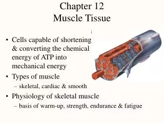
Chapter 12 Muscle Tissue. Cells capable of shortening & converting the chemical energy of ATP into mechanical energy Types of muscle skeletal, cardiac & smooth Physiology of skeletal muscle basis of warm-up, strength, endurance & fatigue. Universal Characteristics of Muscle.

E N D
Chapter 12Muscle Tissue • Cells capable of shortening & converting the chemical energy of ATP into mechanical energy • Types of muscle • skeletal, cardiac & smooth • Physiology of skeletal muscle • basis of warm-up, strength, endurance & fatigue
Universal Characteristics of Muscle • Responsiveness (excitability) • capable of response to chemical signals, stretch or other signals & responding with electrical changes across the plasma membrane • Conductivity • local electrical change triggers a wave of excitation that travels along the muscle fiber • Contractility -- shortens when stimulated • Extensibility -- capable of being stretched • Elasticity -- returns to its original resting length after being stretched
Skeletal Muscle • Voluntary striated muscle attached to one or more bones • Muscle fibers (myofibers) as long as 30 cm • Exhibits alternating light and dark transverse bands or striations • reflects overlapping arrangement of internal contractile proteins • Under conscious control
Series-Elastic Components • Connective tissue elements between muscle fiber and bone or other attachment • endomysium, perimysium, epimysium, fascia, tendon • Not excitable or contractile, but are extensible & elastic • calcaneal tendon produces thrust as toes push off • Behavior during contraction • when a muscle begins to contract, internal tension stretches the series-elastic components • when these are taut, external tension begins to move the load as the muscle cells shorten
Muscle Fibers • Multiple flattened nuclei against inside of plasma membrane • due to fusion of multiple myoblasts during development • unfused satellite cells near can multiply to produce a small number of new myofibers • Sarcolemma has tunnel-like infoldings or transverse (T) tubules that penetrate the cell • carry electric current to cell interior • Sarcoplasm is filled with myofibrils (bundles of parallel protein microfilaments called myofilaments) • glycogen for stored energy & myoglobin binding oxygen • Sarcoplasmic reticulum is series of dilated, calcium storage sacs called terminal cisternae
Thick Filaments • Made of 200 to 500 myosin molecules • 2 entwined golf clubs • Arranged in a bundle with heads directed outward in a spiral array around the bundled tails • heads found on each end with central area a bare zone with no heads
Thin Filaments • Two strands of fibrous (F) actin intertwined • each subunit is a globular (G) actin with an active site • Groove hold tropomyosin molecules blocking the active sites • smaller, calcium-binding troponin molecules stuck to tropomyosin
Elastic Filaments • Huge springy protein called titin (connectin) • runs through core of each thick filament • connects thick filament to Z disc structure • Functions • keep thick & thin filaments aligned • resist overstretching • help the cell recoil to its resting length (elasticity)
Regulatory & Contractile Proteins • Myosin & actin are contractile proteins • they do work of shortening the muscle • Tropomyosin & troponin are regulatory proteins • act like a switch that starts & stops shortening
I A I Striations • Dark A bands (regions) alternating with lighter I bands (regions) • anisotrophic (A) and isotropic (I) stand for the way these regions affect polarized light • A band is thick filament region • lighter, central H band area contains no thin filaments • I band is thin filament region • bisected by Z disc protein anchoring thick & thin • from one Z disc to the next is a sarcomere
Relaxed versus Contracted Sarcomere • Muscle cells shorten because their sarcomeres shorten • pulling Z discs closer together • pulls on sarcolemma • Notice neither thick nor thick filaments change length during shortening • Their overlap changes as sarcomeres shorten
Motor Neurons • Skeletal muscle must be stimulated by a nerve or it will not contract (paralyzed) • Cell bodies of somatic motor neurons are in brainstem or spinal cord • Axons of somatic motor neurons are called somatic motor fibers • each branches, on average, into 200 terminal branches that supply one muscle fiber each • Each motor neuron and all the muscle fibers it innervates are called a motor unit
Motor Units • A motor neuron & the muscle fibers it innervates • dispersed throughout the muscle • when contract together causes weak contraction over wide area • provides ability to sustain long-term contraction as motor units take turns resting (postural control) • Fine control • small motor units contain as few as 20 muscle fibers per nerve fiber • eye muscles • Strength control • gastrocnemius muscle has 1000 fibers per nerve fiber
Neuromuscular Junctions • Synapse is region where nerve fiber makes a functional contact with its target cell (NMJ) • Neurotransmitter released from nerve fiber causes stimulation of muscle cell (acetylcholine) • Components of synapse • synaptic knob is swollen end of nerve fiber • contains vesicles filled with ACh • motor end plate is region of muscle cell surface • has ACh receptors which bind ACh released from nerve • acetylcholinesterase is enzyme that breaks down ACh & causes relaxation • schwann cell envelopes & isolates NMJ
Neuromuscular Toxins & Paralysis • Pesticides contain cholinesterase inhibitors that bind to acetylcholinesterase & prevent it from degrading ACh • spastic paralysis & possible suffocation • Tetanus or lockjaw is spastic paralysis caused by toxin of Clostridium bacteria • blocks glycine release in the spinal cord & causes overstimulation of the muscles • Flaccid paralysis with limp muscles unable to contract caused by curare that competes with ACh
Electrically Excitable Cells (muscle & nerve) • Plasma membrane is polarized or charged • resting membrane potential is due to Na+ outside of cell and K+ & other anions inside of cell • difference in charge across the membrane is potential • inside is slightly more negative (-90 mV) • Plasma membranes exhibit voltage changes in response to stimulation • ion gates open allowing Na+ to rush into cell and then K+ to rush out of cell (quick up-and-down voltage shift is called action potential) • spreads over cell surface as nerve signal or impulse
Muscle Contraction & Relaxation • Four phases involved in this process • excitation where action potentials in the nerve lead to formation of action potentials in muscle fiber • excitation-contraction coupling refers to action potentials on the sarcolemma activate myofilaments • contraction is shortening of muscle fiber or at least formation of tension • relaxation is return of fiber to its resting length • Images will be used to demonstrate the steps of each of these phases
Excitation (steps 1 & 2) • Nerve signal stimulates voltage-gated calcium channels that result in exocytosis of synaptic vesicles containing ACh
Excitation (steps 3 & 4) • Binding of ACh opens Na+ and K+ channels resulting in an end-plate potential (EPP)
Excitation (step 5) • Voltage change in end-plate region (EPP) opens nearby voltage-gated channels in plasma membrane producing an action potential
Excitation-Contraction Coupling(steps 6&7) • Action potential spreading over sarcolemma reaches T tubules -- voltage-gated channels open in T tubules causing calcium gates to open in SR
Excitation-Contraction Coupling(steps 8&9) • Calcium release causes binding of myosin to active sites on actin
Contraction (steps 10 & 11) • Myosin head with an ATP molecule bound to it can form a cross-bridge (myosin ATPase releases the energy allowing the head to move into position)
Contraction (steps 12 & 13) • Power stroke shows myosin head releasing the ADP & phosphate and flexing as it pulls thin filament along -- binding of more ATP releases head from the thin filament
Relaxation (steps 14 & 15) • Stimulation ceases and acetylcholinesterase removes ACh from receptors so stimulation of the muscle cell ceases
Relaxation (step 16) • Active transport pumps calcium back into SR where it binds to calsequestrin • ATP is needed for muscle relaxation as well as muscle contraction
Relaxation (steps 17 & 18) • Loss of calcium from sarcoplasm results in hiding of active sites and cessation of the production or maintenance of tension
Rigor Mortis • Stiffening of the body beginning 3 to 4 hours after death -- peaks at 12 hours after death & diminishes over next 48 hours • Deteriorating sarcoplasmic reticulum releases calcium • Activates myosin-actin cross bridging & muscle contraction • Muscle relaxation requires ATP & ATP production is no longer produced after death • Fibers remain contracted until myofilaments decay
ATP Sources • All muscle contraction depends on ATP • Pathways of ATP synthesis • anaerobic fermentation (ATP production limited) • occurs without oxygen producing toxic lactic acid • aerobic respiration (far more ATP produced) • requires continuous oxygen supply produces H2O & CO2
Cardiac Muscle • In comparison to skeletal muscle, the cells are shorter, thicker, branched and linked to each other at intercalated discs • electrical gap junctions allow cells to stimulate their neighbors & mechanical junctions keep the cells from pulling apart • sarcoplasmic reticulum is less developed but T tubules are larger to admit Ca+2 from extracellular fluid • Autorhythmic due to pacemaker cells • Uses aerobic respiration almost exclusively • large mitochondria make it resistant to fatigue
Smooth Muscle • Fusiform cells with one nucleus • 30 to 200 microns long & 5 to 10 microns wide • no visible striations, sarcomeres or Z discs • thin filaments attach to dense bodies scattered throughout sarcoplasm & on sarcolemma • SR is scanty & has no T tubules • calcium for contraction comes from extracellular fluid • Nerve supply is autonomic, if any is present
Types of Smooth Muscle • Multiunit smooth muscle • in largest arteries, iris, pulmonary air passages, arrector pili muscles • terminal nerve branches synapse on individual myocytes in a motor unit • independent contraction • Single-unit smooth muscle • in most blood vessels & viscera as circular & longitudinal muscle layers • electrically coupled by gap junctions • large number of cells contract as a unit
Stimulation of Smooth Muscle • Involuntary & contracts without nerve stimulation • hormones, CO2, low pH, stretch, O2 deficiency • pacemaker cells in GI tract are autorhythmic • Autonomic nerve fibers have beadlike swellings called varicosities containing synaptic vesicles • stimulates multiple myocytes at diffuse junctions
Features of Contraction and Relaxation • Calcium triggering contraction is extracellular • enters cell through channels triggered by voltage, hormones, neurotransmitters or stretching of the cell • calcium ion binds to calmodulin -- activates myosin light-chain kinase which activates the myosin head with ATP to bind actin -- power stroke occurs when hydrolyzes 2nd ATP • Thin filaments pull on intermediate filaments attached to dense bodies on the plasma membrane • shortens the entire cell in a twisting fashion • Contraction & relaxation very slow in comparison • slow myosin ATPase enzyme & slow pumps that remove Ca+2 • Uses l0-300 times less ATP to maintain the same tension • latch-bridge mechanism maintains tetanus (muscle tone) • keeps arteries in state of partial contraction (vasomotor tone)
Responses to Stretch • Stretch opens mechanically-gated calcium channels causing muscle response • food entering the esophagus brings on peristalsis • Stress-relaxation response necessary for hollow organs that gradually fill (urinary bladder) • when stretched, tissue briefly contracts then relaxes • Must contract forcefully when greatly stretched • thick filaments have heads along their entire length • no orderly filament arrangement -- no Z discs • Plasticity is ability to adjust tension to degree of stretch such as empty bladder is not flabby
Muscular Dystrophy • Group of hereditary diseases in which skeletal muscles degenerate & are replaced with adipose • Mainly a disease of males • appears as child begins to walk • rarely live past 20 years of age • Normal allele makes dystrophin, a protein that links actin filaments to cell membrane • absence of dystrophin leads to torn cell membranes • Fascioscapulohumeral MD -- facial & shoulder muscle only
Myasthenia Gravis • Autoimmune disease where antibodies attack NMJ and bind ACh receptors together in clusters • fibers remove the receptors • less and less sensitive to ACh • drooping eyelids and double vision • difficulty swallowing • weakness of the limbs • respiratory failure • Disease of women between ages of 20 and 40 • Treated with cholinesterase inhibitors, thymus removal or immunosuppressive agents