Download
1 / 163
1.63k likes | 1.7k Views
This lecture covers the types and functions of muscle tissue, along with the structure and components of muscle fibers. Topics include isotonic and isometric contractions, neuromuscular junctions, and the anatomy of cardiac and smooth muscles. Presented by Zagipa Sapakhova, PhD, Associate Professor at Shakarim State University of Semey.

E N D
SHAKARIM STATE UNIVERSITY OF SEMEY Lecture 11-12Muscle Tissue Sapakhova Zagipa, PhD, associated professor
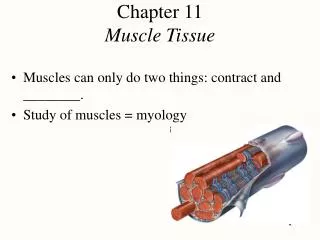
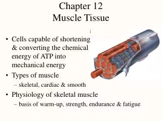
Muscular Tissue • Alternating contraction and relaxation of cells • Chemical energy changed into mechanical energy
3 Types of Muscle Tissue • Skeletal muscle • attaches to bone, skin or fascia • striated with light & dark bands visible with scope • voluntary control of contraction & relaxation
3 Types of Muscle Tissue • Cardiac muscle • striated in appearance • involuntary control • autorhythmic because of built in pacemaker
3 Types of Muscle Tissue • Smooth muscle • attached to hair follicles in skin • in walls of hollow organs -- blood vessels & GI • nonstriated in appearance • involuntary
Functions of Muscle Tissue • Producing body movements • Stabilizing body positions • Regulating organ volumes • bands of smooth muscle called sphincters • Movement of substances within the body • blood, lymph, urine, air, food and fluids, sperm • Producing heat • involuntary contractions of skeletal muscle (shivering)
Muscle Fiber or Myofibers • Muscle cells are long, cylindrical & multinucleated • Sarcolemma = muscle cell membrane • Sarcoplasm filled with tiny threads called myofibrils & myoglobin (red-colored, oxygen-binding protein)
Transverse Tubules • T (transverse) tubules are invaginations of the sarcolemma into the center of the cell • filled with extracellular fluid • carry muscle action potentials down into cell • Mitochondria lie in rows throughout the cell • near the muscle proteins that use ATP during contraction
Myofibrils & Myofilaments • Muscle fibers are filled with threads called myofibrils separated by SR (sarcoplasmic reticulum) • Myofilaments (thick & thin filaments) are the contractile proteins of muscle
Sarcoplasmic Reticulum (SR) • System of tubular sacs similar to smooth ER in nonmuscle cells • Stores Ca+2 in a relaxed muscle • Release of Ca+2 triggers muscle contraction
Filaments and the Sarcomere • Thick and thin filaments overlap each other in a pattern that creates striations (light I bands and dark A bands) • They are arranged in compartments called sarcomeres, separated by Z discs. • In the overlap region, six thin filaments surround each thick filament
Rigor Mortis • Rigor mortis is a state of muscular rigidity that begins 3-4 hours after death and lasts about 24 hours • After death, Ca+2 ions leak out of the SR and allow myosin heads to bind to actin • Since ATP synthesis has ceased, crossbridges cannot detach from actin until proteolytic enzymes begin to digest the decomposing cells.
Neuromuscular Junction (NMJ) or Synapse • NMJ = myoneural junction • end of axon nears the surface of a muscle fiber at its motor end plate region (remain separated by synaptic cleft or gap)
Structures of NMJ Region • Synaptic end bulbs are swellings of axon terminals • End bulbs contain synaptic vesicles filled with acetylcholine (ACh) • Motor end plate membrane contains 30 million ACh receptors.
Events Occurring After a Nerve Signal • Arrival of nerve impulse at nerve terminal causes release of ACh from synaptic vesicles • ACh binds to receptors on muscle motor end plate opening the gated ion channels so that Na+ can rush into the muscle cell • Inside of muscle cell becomes more positive, triggering a muscle action potential that travels over the cell and down the T tubules • The release of Ca+2 from the SR is triggered and the muscle cell will shorten & generate force • Acetylcholinesterase breaks down the ACh attached to the receptors on the motor end plate so the muscle action potential will cease and the muscle cell will relax.
Isotonic and Isometric Contraction • Isotonic contractions = a load is moved • concentric contraction = a muscle shortens to produce force and movement • eccentric contractions = a muscle lengthens while maintaining force and movement • Isometric contraction = no movement occurs • tension is generated without muscle shortening • maintaining posture & supports objects in a fixed position
Anatomy of Cardiac Muscle • Striated , short, quadrangular-shaped, branching fibers • Single centrally located nucleus • Cells connected by intercalated discs with gap junctions • Same arrangement of thick & thin filaments as skeletal
Appearance of Cardiac Muscle • Striated muscle containing thick & thin filaments • T tubules located at Z discs & less SR
Microscopic Anatomy of Smooth Muscle • Small, involuntary muscle cell -- tapering at ends • Single, oval, centrally located nucleus • Lack T tubules & have little SR for Ca+2 storage
Microscopic Anatomy of Smooth Muscle • Thick & thin myofilaments not orderly arranged so lacks sarcomeres • Sliding of thick & thin filaments generates tension • Transferred to intermediate filaments & dense bodies attached to sarcolemma • Muscle fiber contracts and twists into a helix as it shortens -- relaxes by untwisting
Muscle Tissue • One of 4 primary tissue types, divided into: • skeletal muscle • cardiac muscle • smooth muscle Without these muscles, nothing in the body would move and no body movement would occur
Skeletal Muscles- Organs of skeletal muscle tissue - are attached to the skeletal system and allow us to move • Muscular System- Includes only skeletal muscles Skeletal Muscle Structures • Muscle tissue (muscle cells or fibers) • Connective tissues • Nerves • Blood vessels
6 Functions of Skeletal Muscles • Produce skeletal movement • Maintain body position and posture • Support soft tissues • Guard body openings (entrance/exit) • Maintain body temperature • Store Nutrient reserves
How is muscle tissue organized at the tissue level?Organization of Connective Tissues Figure 10–1
Organization of Connective Tissues • Muscles have 3 layers of connective tissues: 1. Epimysium-Exterior collagen layer • Connected to deep fascia • Separates muscle from surrounding tissue 2. perimysium- Surrounds muscle fiber bundles (fascicles) • Contains blood vessel and nerve supply to fascicles 3. endomysium
3. Endomysium • Surrounds individual muscle cells (muscle fibers) • Contains capillaries and nerve fibers contacting muscle cells • Contains satellite cells (stem cells) that repair damage
Muscle Attachments • Endomysium, perimysium, and epimysium come together: • at ends of muscles • to form connective tissue attachment to bone matrix • i.e.,tendon (bundle) or aponeurosis (sheet)
NervesSkeletal muscles are voluntary muscles, controlled by nerves of the central nervous system Blood Vessels • Muscles have extensive vascular systems that: • supply large amounts of oxygen • supply nutrients • carry away wastes
What are the characteristics of skeletal muscle fibers? • Skeletal muscle cells are called fibers Figure 10–2
Skeletal Muscle Fibers • Are very long • Develop through fusion of mesodermal cells (myoblasts- embryonic cells)) • Become very large • Contain hundreds of nuclei –multinucleate • Unfused cells are satellite cells- assist in repair after injury
Organization of Skeletal Muscle Fibers Figure 10–3
The Sarcolemma • The cell membrane of a muscle cell • Surrounds the sarcoplasm (cytoplasm of muscle fiber) • A change in transmembrane potential begins contractions • All regions of the cell must contract simultaneously
Transverse Tubules (T tubules) • Transmit action potential – impulses through cell • Allow entire muscle fiber to contract simultaneously • Have same properties as sarcolemma • Filled with extracellular fluid
Myofibrils- 1-2um in diameter • Lengthwise subdivisions within muscle fiber • Made up of bundles of protein filaments (myofilaments) • Myofilaments - are responsible for muscle contraction 2 Types of Myofilaments • Thin filaments: • made of the protein actin • Thick filaments: • made of the protein myosin
Sarcoplasmic Reticulum (SR) • A membranous structure surrounding each myofibril • Helps transmit action potential to myofibril • Similar in structure to smooth endoplasmic reticulum • Forms chambers (terminal cisternae) attached to T tubules
A Triad • Is formed by 1 T tubule and 2 terminal cisterna Cisternae • Concentrate Ca2+ (via ion pumps) • Release Ca2+ into sarcomeres to begin muscle contraction
Structural components of the Sarcomeres -The contractile units of muscle -Structural units of myofibrils -Form visible patterns within myofibrils Figure 10–4
Muscle Striations • A striped or striated pattern within myofibrils: • alternating dark, thick filaments(A bands) and light, thin filaments(I bands)
M Lines and Z Lines • M line: • the center of the A band • at midline of sarcomere • Z lines: • the centers of the I bands • at 2 ends of sarcomere Zone of Overlap • The densest, darkest area on a light micrograph • Where thick and thin filaments overlap
The H Zone • The area around the M line • Has thick filaments but no thin filaments Titin • Are strands of protein • Reach from tips of thick filaments to the Z line • Stabilize the filaments
Sarcomere Structure Figure 10–5
Sarcomere Function • Transverse tubules encircle the sarcomere near zones of overlap • Ca2+ released by SR causes thin and thick filaments to interact
Level 1: Skeletal Muscle Level 2: Muscle Fascicle Figure 10–6 (1 of 5)
Level 3: Muscle Fiber Level 4: Myofibril Figure 10–6 (3 of 5)