Download
1 / 44
1.05k likes | 4.19k Views
Secondary Structure Motifs of Proteins . Chapter 2. Their Diverse Functions Require Proteins to Have Irregular Structures.

E N D
Secondary StructureMotifs of Proteins Chapter 2
Their Diverse Functions Require Proteins to Have Irregular Structures • Kendrew's model of the low-resolution structure of myoglobin shown in three different views. The sausage-shaped regions represent helices, which are arranged in a seemingly irregular manner to form a compact globular molecule. (Courtesy of J.C. Kendrew.)
Types of Secondary Structure • There are three common secondary structures in proteins, namely alpha helices, beta sheets, and turns. • That which cannot be classified as one of the standard three classes is usually grouped into a category called "other" or "random coil". This designation is unfortunate as no portion of a protein’s three dimensional structure is truly random and it is usually not a coil. • A common element of most secondary structures is the presence of characteristic hydrogen bonds e.g., C=O of residue i to HN of residue i+4 (i, i+4). They are formed when a number of consecutive residues have the same phi and psi angles.
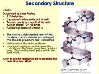
Helix • In a helical conformation, the relationship of one peptide unit to the next is the same for all alpha-carbons. This means that the dihedral angle pairs phi and psi (phii, psii) are the same for each residue in the helical conformation. • Helices are classified as repetitive secondary structure since their backbone phi and psi angles repeat • Two parameters describe the helix about this axis: n - the number of residues per helical turn r - the rise per helical residue • By convention, a positive value of n denotes a right-handed helix. (Curling the fingers of your right hand along the helical path, your thumb will point in the direction of your fingertips if the helix is right-handed.)
Three Regular Polypeptide Helices -helix a-helix 310 helix phi psi a-helix -57.8 -47.0 310-helix -74.0 -4.0 p-helix -57.1 -69.7 Idealized model of the conformations of polyalanine are displayed.
The Alpha (a) Helix • Main-chain N and O atoms are hydrogen-bonded to each other helices. There are 3.6 residues per turn in an helix, which corresponds to 5.4 Å (1.5 Å per residue).
The Alpha (a) Helix The Side chains project out from the alpha helix .
The Alpha Helix has a Dipole Moment Negatively charged groups such as phosphate ions frequently bind to the amino ends of a helices. The dipole moment of an a helix as well as the possibility of hydrogen-bonding to free NH groups at the end of the helix favors such binding. • (a) The dipole of a peptide unit. Values in boxes give the approximate fractional charges of the atoms of the peptide unit. (b) The dipoles of peptide units are aligned along the a-helical axis, which creates an overall dipole moment in the a helix, positive at the amino end and negative at the carboxyl end.
The Helical Wheels • The helical wheel or spiral. Amino acid residues are plotted every (360/3.6) 100° around the spiral. • Green is an amino acid with a hydrophobic side chain, blue is a polar side chain, and red is a charged side chain.
Some Amino Acids are Preferred in a Helix • Eight Most Common Residues as Helix Formers: • Glu, Met, Ala, Leu, Lys, Phe, Gln, Trp • Eight Least Common Residues as Helix Formers: • Gly, Pro, Asn, Tyr, Cys, Ser, Thr, Arg
310 Helix • Only 3.4 % of the residues are involved in 310 helices in the Kabsch and Sander database (1983), and nearly all those in helical segments containing 1 - 3 hydrogen bonds (96 % ± 4 residues). • The average of the backbone dihedral angles were found to differ slightly from the ideal 310 helix (-74.0, -4.0) with values of -71.0 and -18.0 degrees, for phi and for psi, respectively. It has a larger radius (2.0 versus 1.9 Å) and a larger number of residues per helical turn (3.2 versus 3.0). • The end result being a slightly better staggering of sidechains along the helical axis. • Hydrogen bonds within a 310-helix also display a repeating pattern in which the backbone C=O of residue i hydrogen bonds to the backbone HN of residue i+3.
-helix • Hydrogen bonds within a p-helix display a repeating pattern, in which the backbone C=O of residue i hydrogen bonds to the backbone HN of residue i+5. • The p-helix is an extremely rare secondary structural element in proteins. • The infrequency of this particular form of secondary structure stems from the following properties: 1. the phi and psi angles lie at the very edge of an allowed, minimum energy region of the Ramachandran (phi, psi) map. 2. the p-helix requires that the angle tau (N-CA-C') be larger (114.9) than the standard tetrahedral angle of 109.5 degrees. 3. the large radius of the p-helix forms an axial hole too small for solvent water to fill. 4. side chains are more staggered than the ideal 310 helix but not as well as the -helix.
b-sheet • Beta sheets are another major structural element in globular proteins containing 20 – 28 % of all residues (Kabsch & Sander, 1983; Creighton, 1993). • The basic unit of a beta sheet is a beta strand with approximate backbone dihedral angles phi = -120 and psi = +120 producing a translation of 3.2 to 3.4 Å/residue for residues in anti-parallel and parallel strands, respectively. • Due to the extended nature of the chain, there are no significant intra-segment hydrogen bonds and van der Waals interactions between atoms of neighboring residues. This extended conformation is only stable as part of a beta sheet where contributions from hydrogen bonds and van der Waals interactions between aligned strands exert a stabilizing influence. • The beta sheet is sometimes called the beta "pleated" sheet since sequentially neighboring Ca atoms are alternately above and below the plane of the sheet.
Main-chain NH and O atoms within a b sheet are hydrogen bonded to each other. The amino acids in successive strands have alternating directions (anti-parallel). Anti-parallel b sheet
Anti-parallel b sheet A residue in an antiparallel beta strand has values of -139 and +135 degrees for the backbone dihedral angles phi and psi,respectively. Antiparallel beta sheets are thought to be intrinsically more stable than parallel sheets due to the more optimal orientation of the interstrand hydrogen bonds and that peptide bond dipoles of nearest neighbors within a strand cancel whereas in the parallel sheet, components of the dipoles parallel to the strands align and may interact unfavorably.
The amino acids in the aligned strands run in the same direction. Parallel b sheet
Twist of b Sheet • The classical beta sheets originally proposed are planar but most sheets observed in globular proteins are twisted (0 to 30 º/ residue). • Antiparallel beta sheets are more often twisted than parallel sheets. This twist is always of the same handedness, but unfortunately, it has been described using two conflicting conventions in the literature. If defined in terms of the progressive twist of the hydrogen-bonding direction, the twist is right-handed. • Two-stranded beta strands show the largest twists.
Another irregularity found in antiparallel beta sheets is the hydrogen-bonding of two residues from one strand with one residue from the other called a beta bulge. Bulges are most often found in antiparallel sheets with ~5 % of bulges occurring in parallel strands (Richardson, 1981). Bulges, like "Turns" effect the directionality of the polypeptide chain. b-Bulge
Turns • Turns are the third of the three "classical" secondary structures. Approximately one-third of all residues in globular proteins are contained in turns that serve to reverse the direction of the polypeptide chain. • This is perhaps not so surprising since the diameter of the average globular protein domain is roughly 25 Å (an extended polypeptide conformation would require ~7 residues to traverse the domain before having to change directions). • Turns are located primarily on the protein surface and accordingly contain polar and charged residues. Antibody recognition, phosphorylation, glycosylation, hydroxylation, and intron/exon splicing are found frequently at or adjacent to turns.
The hydrogen bond between CO of residue i and NH of residue i+2. The dihedral angles of residue i+1 are (70, -60) and (-70, 60) for phi and psi of the classical and inverse gamma turns. Gamma Turn
Type I Turn. • The hydrogen bond between CO of residue i and NH of residue i+3. • The backbone dihedral angles are (-60, -30) and (-90, 0) of residues i+1 and i+2, respectively, for the type I turn. • Proline is often found in position i+1 in type I turns as its phi angle is restricted to -60 and its imide nitrogen does not require a hydrogen bond. Glycine is favored in this position in the type II' as it requires a positive (left-handed) phi value.
Type II Turn. • The hydrogen bond between CO of residue i and NH of residue i+3. • The backbone dihedral angles are (-60, 120) and (80, 0) of residues i+1 and i+2, respectively, for the type II turn. • Glycine is favored in this position in the type II' as it requires a positive (left-handed) phi value.
Type III Turn. • The hydrogen bond between CO of residue i and NH of residue i+3. • This is a single turn of right-handed (III) and left-handed (III') 310 helix. The backbone dihedral angles are (-60, -30) and (-60, -30) of residues i+1 and i+2, respectively, for the classical type III turn.
Eight most common residues for beta-sheet Val, Ile, Tyr, Trp, Phe, Leu, Cys, Thr Eight least common residues for beta-sheet Glu, Asp, Pro, Ser, Lys, Gly, Ala, Asn Eight most common residues for turns Gly, Asn, Pro, Asp, Ser, Cys, Tyr, Lys Eight least common residues for turns Ile, Val, Met, Leu, Phe, Ala, Glu, Trp Preferred Residues for b Sheet and Turns
Loops • In Leszczynski & Rose (1986), out of 67 proteins surveyed, they tabulated 26 % helix, 19% sheet, 26 % turns and 21 % in loops. • These loop structures contain between 6 and 16 residues and are compact and globular in structure. Like turns, they generally contain polar residues and hence are predominantly at the protein surface.
Adjacent antiparallel b strands are joined by hairpin loops. Such loops are frequently short and do not have regular secondary structure. Nevertheless, many loop regions in different proteins have similar structures. b-hairpin Loop
Richardson Diagrams Myoglobin Triosephosphate isomerase Cylinder for a helices; arrows for b strands, which gives the direction of the strand from N to C; and the ribbons for the remaining part.
Beta Sheet Topology Diagrams • Beta sheets are usually represented simply by arrows in topology diagrams that show both the direction of each strand and the way the strands are connected to each other along the polypeptide chain. plastocyanin transcarbamoylase flavodoxin
Super Secondary Structures (Motifs) • Simple combinations of a few secondary structure elements with a specific geometric arrangement are called super secondary structures or motifs. • They may have functional and structural significance. • Common motifs: • Helix-turn-helix b-hairpin, b-meander b-barrel, Geek key bab
Helix-Turn-Helix Motif • Two helices that are connected by a short loop region in a specific geometric arrangement constitute a helix-turn-helix motif. (a) the DNA-binding motif and (b) the calcium-binding motif, which are present in many proteins whose function is regulated by calcium.
EF-hand Calcium-binding Motif • The calcium atom is bound to one of the motifs in the muscle protein troponin-C through six oxygen atoms: one each from the side chains of Asp (D) 9, Asn (N) 11, and Asp (D) 13; one from the main chain of residue 15; and two from the side chain of Glu (E) 20. In addition, a water molecule (W) is bound to the calcium atom.
Amino Acid Sequences of EF-hand Motifs 1 3 5 7 9 12 The side chains of hydrophobic residues on the flanking helices form a hydrophobic core between the a helices
The b Hairpin Motif • The hairpin motif is very frequent in b sheets and is built up from two adjacent b strands that are joined by a loop region. Snake Venom Erabutoxin Bovine Trypsin Inhibitor
Greek Key Motif • The Greek key motif is found in antiparallel b sheets when four adjacent b strands are arranged in the pattern shown as a topology diagram in (a). The three dimensional structure of the enzyme Staphylococcus Nuclease shown in (b) in blue and red is also a Greek key motif.
Forming Greek Key Motif • Suggested folding pathway from a hairpin-like structure to the Greek key motif. • Beta strands 2 and 3 fold over such that strand 2 is aligned adjacent and antiparallel to strand 1.
b-a-b Motif • Two adjacent parallel b strands are usually connected by an a helix from the C-terminus of strand 1 to the N-terminus of strand 2. • Most protein structures that contain parallel b sheets are built up from combinations of such b-a-b motifs.
b-a-b Handedness • The b-a-b motif can in principle have two "hands." • (a) This connection with the helix above the sheet is found in almost all proteins and is called right-handed because it has the same hand as a right-handed a helix. • (b) The left-handed connection with the helix below the sheet.
Domain Organization • Small protein molecules like the epidermal growth factor, EGF, are comprised of only one domain. Others, like the serine proteinase chymotrypsin, are arranged in two domains that are required to form a functional unit. Many of the proteins that are involved in blood coagulation and fibrinolysis have long polypeptide chains that comprise different combinations of domains.
Domains • "Within a single subunit [polypeptide chain], contiguous portions of the polypeptide chain frequently fold into compact, local semi-independent units called domains." - Richardson, 1981 • Domains may be considered to be connected units, which are to varying extents independent in terms of their structure, function and folding behavior. • Each domain can be described by its fold. While some proteins consist of a single domain, others consist of several or many. A number of globular protein chains consist of two or three domains appearing as 'lobes'. • In other cases, the domains may be of a very different nature. For example, some proteins located in cell membranes have a globular intracellular or extracellular domain distinct from that which spans the membrane.
Adjacent Motifs Motifs that are adjacent in the amino acid sequence are also usually adjacent in the three-dimensional structure. Triose-phosphate isomerase is built up from four b-a-b-a motifs that are consecutive both in the amino acid sequence (a) and in the three dimensional structure (b).
Mosaic Proteins • Mosaic proteins are those which consist of many repeated copies of one or a few domains, all within one polypeptide chain. • Many extracellular proteins are of this nature. The domains in question are termed modules and are sometimes relatively small. Note that this term is often applied to sequences whose structures may not be known for certain.