Download

1 / 24
280 likes | 581 Views
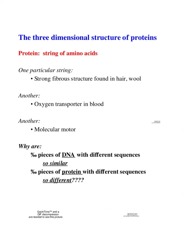
Structure of Proteins. 3D structure determined by amino acid sequence Structure - Function Native structure of a protein = functionally, folded conformation Protein conformation stabilized by 1. disulfide bonds 2. weak noncovalent interactions ( H-bonds, hydrophobic & ionic ).

E N D
Structure of Proteins 3D structure determined by amino acid sequence Structure - Function Native structure of a protein = functionally, folded conformation Protein conformation stabilized by 1. disulfide bonds 2. weak noncovalent interactions (H-bonds, hydrophobic & ionic) Chymotrypin Glycine
3D Structure of Proteins Primary structure = amino acids linked together Peptide bond is rigid and planar
Secondary Structure of Proteins Important elements - steric clashes & H-bonding Basic types of secondary structure: Helices, Sheets, Turns and Coils Helices H-bond
Secondary Structure of Proteins Helices Ionic interaction between R groups of AAs three residues apart Arg Asp
Secondary Structure of Proteins sheets Backbone is extended into a zigzag structure Arranged side-by-side to form a structure (pleats) Important Forces = H-bonds and steric clash Layering of >2 sheets R groups must be small (Gly, Ala)
Secondary Structure of Proteins turns Occur frequently in globular proteins, 180˚ turn involving 4 Aas Used to: 1. Reverse direction of polypeptide chain 2. Connect helices/ sheets and within sheets Important forces: Amino acids used: Gly - because it is small and flexible Pro - because of cis conformation of peptide bond forms a tight turn
Tertiary Structure Overall 3D arrangement of all atoms in a protein Long range contacts between AAs in a single polypeptide chain
Quarternary Structure Long range contacts between AAs in a different polypeptide chain
Fibrous Proteins Mainly structural role
Fibrous Proteins -Keratins Found in: mammals, provide strength Hair, wool, nails, claws, quills, horns, hooves, skin Strengthened by: Disulfide bonds
Fibrous Proteins -Keratins Permanent waving of hair 1. Reduce disulfide bonds 2. Moist heat breaks H-bonds and causes uncoiling of helix 3. Remove reducing agent, add oxidizing agent, new S-S bonds
Fibrous Proteins • Collagen helices, left-handed helix with 3 amino acids per turn 35% Gly, 11% Ala, 21% Pro/4-Hyp (Gly-X-Y) repeat with X as Pro and Y as 4-Hyp • Coiled-coil, three separate polypeptides called chains are supertwisted • Provide strength (stronger than ??) • Connective tissue (tendons, cartilage, organic matrix of bone, cornea)
Fibrous Proteins Collagen Rigid and brittle bones caused by: Crosslinks in collagen fibrils over time Gly-X-Y repeat important - single change results in disease Osteogenesis imperfecta - abnormal bone formation in babies Ehlers-Danlos syndrome - loose joints Both diseases involve: mutation of Gly to a different amino acid
Fibrous Proteins Silk Fibrous protein of silk = Fibroin Secondary structure present: sheets Forces involved: H-bonds between different sheets Made by: insects and spiders Silk does not stretch because it is already highly extended
Globular Proteins helices and sheets and turns and ……… noncovalent interactions Arrangement of different secondary structural elements: Compact conformation Folding provides structural diversity Globular proteins = enzymes, transport proteins, motor proteins, regulatory proteins, immunoglobulins, etc. First understanding of globular proteins came from: x-ray structure of myoglobin (oxygen-binding protein in muscle) Iron protoporphyrin (heme) Single polypeptide chain helix turn
Globular Proteins Other important forces in globular proteins: Hydrophobic interactions Hydrophobic aa
Globular Proteins Well-studied example: Myoglobin Flat heme group rests in crevice of protein
Globular Proteins Variety of Tertiary Structures Disulfide bond Heme Disulfide bond Respiratory chain in mitochondria Egg white and human tears Cleaves polysaccharides Enzyme secreted by pancreas Hydrolyzes RNA
Protein Denaturation & Folding AA sequence determines tertiary structure Importance of native structure Loss of structure = loss of function
Protein Denaturation & Folding Rapid stepwise folding
Protein Denaturation & Folding Defects in folding may lead to disease AA mutation in CFTR - cystic fibrosis BUT No AA mutation (except in inherited forms) just misfolding in (PrP) Prion Protein
Protein Denaturation & Folding Proteins undergo assisted folding “molecular chaperones” assist in folding