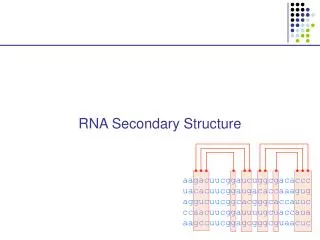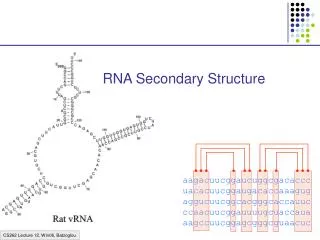
Secondary Structure
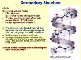
Secondary Structure. a helix Discovered by Linus Pauling 2 Nobel prizes. Discovered folding while sick in bed! Treated poorly as a result of his anti-nuclear stands - 2 nd NB prize Pushed high doses of vitamin C

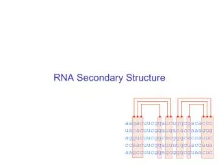
Secondary Structure
E N D
Presentation Transcript
Secondary Structure • ahelix • Discovered by Linus Pauling • 2 Nobel prizes. • Discovered folding while sick in bed! • Treated poorly as a result of his anti-nuclear stands - 2nd NB prize • Pushed high doses of vitamin C • The helix is a right handed twist of the backbone - notice when we are looking at this the side groups are NOT considered • Notice where the amino acids are. • Hydrogen bonding occurs between the carbonyl and the amino group four residues away. The bonding takes place within the same chain. • A run of proline residues lead to breaking the helix structure. Why?
And there are always exceptions! • Proline –Cyclic side chain limits the range of rotation • It is the most conformationally restricted amino acid residue • Glycine- The only amino acid without a beta carbon atom is the smallest of the amino acids • Has almost unlimited rotational freedom and can fit into almost any conformation
Specifics on helical descriptions: • Pitch:the vertical distance per turn. Think of a screw, if you turned a screw one turn this is how far the screw would penetrate • n = residues per turn. This depends on the helix. If it is stretched out (n=2) if it is compressed (n=12) the most stable is 3.6 or nearly 4 amino acids per turn. This is where the best H bonding can occur. (think of the alignment of the donor and acceptor • nth residue:This means any residue in the helix as a reference point • nth + 4 residue:Indicates the residue 4 positions in with n taken as the first. The +sign does not mean add 4 residues to n, just you are looking at a residue 4 amino acids away from the nth residue. • nm:n refers to the residue position, m is the number of atoms in the loop formed by hydrogen bonding. The a helix has an nm of 413 m
b Pleated sheet • Formed when the peptide chain is lined up side by side. This is an extended formation of the backbone • The structure is stabilized by hydrogen bonds between two different chains • -C=O and -NH • Same donors and acceptors as in the helix but not within the same chain Each strand of the b pleated sheet is from 6 to 15 residues
2 arrangements of sheets parallel and anti parallel. Look at the N to C terminus directions
2 arrangements of sheets parallel and anti parallel. Look at the N to C terminus directions • Anti parallel conformations are stronger - alignment of H bonding. • Often found in silk • R groups can interact - glycine and alanine • H bonding within 3 residues can disrupt the sheets - causes bend or turn in the chain.
Unique structure of collagen • A special helical protein • biological significance - fibrous, structural component • Type I collagen is found in bone, tendon and skin, II in cartilage and III in blood vessels • very different amino acid sequence from alpha helix • 3 residues / turn • 1/3 amino acids are glycine Gly X Y
Unique structure of collagen • A special helical protein • Glycine R group face inside others outside • up to 30% are proline or hydroxyproline - important for maintaining secondary structure • hydroxyprolines involved in H bonding of three strands together • helical structure formed by three left handed helices twisted to form a right handed superhelix (gives strength) • hydrogen bonding between 3 helices (thus the glycine) • covalent bonding of lysine between strands necessary for strength
Unique structure of collagen • A special helical protein • Hydroxylations on pro are performed by an enzyme called prolyl hydroxylase, which is an enzyme that requires vitamin C as a cofactor in the reaction. • Absence of vitamin C in the diet reduces hydroxylation of pro, and collagen fibres begin to break down and new collagen not formed properly. • Lack of vitamin C causes scurvy because collagen fibres are not formed properly, and this causes skin lesions, weakened gums so teeth fall out etc.
Unique structure of collagen • A special helical protein • Equally important is hydroxy-lys catalysed by lysine hydroxylase. Attached to the lys residues are three sugars gal-gal-glu, and these enable H-bonding to occur between triple helices, which is essential for stability of the greater complex that binds fibers together to form a matrix bed to binds cells to the matrix and form a tissue.
Collagen Related Disease • Loss of flexibility with age is likely due to increased amount cross-linked collagen compared to younger tissue • Scurvy – problems with sea voyages, lack of food other than salted meats.
Collagen Related Disease • Loss of flexibility with age is likely due to increased amount cross-linked collagen compared to younger tissue • Scurvy – problems with sea voyages, lack of food other than salted meats. • Symptoms include, swollen gums, loose teeth, small black-and-blue spots on the skin, and bleeding from small blood vessels are among the characteristic signs of scurvy. • Caused when vitamin C (ascorbic acid) is lost from diet • Vit C is needed to keep Iron reduced in the active site of prolyl hydroxylase. This is the enzyme responsible for conversion of proline to hydroxyproline. The H bonding of hydroxyproline is vital for the connective protein’s function • In 1795, the British Royal Navy provided a daily ration of lime or lemon juice to all its men. English sailors to this day are called "limeys", for lime was the term used at the time for both lemons and limes.
Collagen Related Disease • Several heritable diseases result from mutations in the collagen • Brittle Bone Disease – results from a Gly-Ala mutation – Consider the consequences of this mutation, both in the protein’s triple helix and the strength of the bone!
Collagen Related Disease • Several heritable diseases result from mutations in the collagen Marfan’s Syndrom and Ehler’s-Danlos syndromes - inherited disorder of connective tissue which affects many organ systems, including the skeleton, lungs, eyes, heart and blood vessels. All resulting from various mutation in collagen and other fibril associated proteins, ultimately affecting the structure and molecular interaction.
Tertiary structure • overall final 3 D shape of a protein assumes • aa side chain interactions responsible for 3 D structures • hydrophobic interactions • salt bridges • hydrogen bonds (know donors and acceptors) • sulfhydryl bonds
Tertiary structure • overall final 3 D shape of a protein assumes • aa side chain interactions responsible for 3 D structures • hydrophobic interactions • salt bridges • hydrogen bonds (know donors and acceptors) • sulfhydryl bonds • Domain - an amino segment of folded protein showing conformational integrity. Or…
Tertiary structure • overall final 3 D shape of a protein assumes • aa side chain interactions responsible for 3 D structures • hydrophobic interactions • salt bridges • hydrogen bonds (know donors and acceptors) • sulfhydryl bonds • Domain - an amino segment of folded protein showing conformational integrity. Or… • A region of the protein that has its own identifiable function. There are dozens of domains identified. A protein can have more than one domain. • Can be made of the whole protein or just part of the protein. • Often coded by their own section of DNA (exon)
Quaternary structure • Proteins with 2 or more peptide chains or subunits • Proteins with 2 or more peptide chains or subunits can be different or identical subunits • loss of quaternary or tertiary (native) structure is called denaturation. Examples include - Heat – to unravel the folding by adding energy – eg. egg whites - Detergent – interfere with hydrophobic interactions - Changes in pH - ionization of R groups • Reducing agents – chemicals which break sulfhydryl bonds by changing the redox state of cys amino acids.
Mad Cow Disease • All known prion diseases are fatal. Since the immune system does not recognize prions as foreign, no natural protection develops. Scrapie in sheep was first described during the18th century. It has been transmitted to other animals such as mink and cats, and more recently to cows (mad cow disease or bovine spongiform encephalopathy, BSE) through contaminated feedstuff. • In New Guinea, the Fore-people contracted kuru by eating the brains of deceased people. Creutzfeldt-Jakob Disease (CJD) frequently arises spontaneously, while fatal familial insomnia (FFI) Gerstmann-Sträussler-Scheinker GSS) disease, and 10-15% of CJD are caused by mutations in the gene encoding the prion protein. A new variant CJD, diagnosed in some 20 patients, may have arisen through transmission of BSE to humans. The Nobel Foundatioin
BSE - Mad Cow; A protein gone wrong • The prion protein exists in two forms. The normal, protein (PrPc) can change its shape to a harmful, disease-causing form (PrPSc). The conversion from PrPc to PrPSc then proceeds via a chain-reaction. When enough PrPSc proteins have been made they form long filamentous aggregates that gradually damage neuronal tissue. The harmful PrPSc form is very resistant to high temperatures, UV-irradiation and strong degradative enzymes.
Prions and protein folding Cerebral cortex -loss of memory and mental acuity (CJD). Thalamus Damage results in insomnia (FFI). Cerebellum Damage results in problems to coordinate body movements and difficulties to walk (kuru, GSS). Brain stem In the mad cow disease (BSE). • Prions affect different regions of the brain. A sponge-like appearance results when nerve cells die. Symptoms depend on which region of the brain is affected. • A precise diagnosis of a prion disease can only be made upon autopsy. The figures show thin sections of diseased brains.FFI, with typical proliferation of astrocytes, the support cells of the brain, is shown to the left (arrows).CJD, with the characteristic spongiform appearance with vacuoles (arrows) is shown to the right.
Prion diseases arise in three different ways • 1. Through horizontal transmission from e.g. a sheep to a cow (BSE). • 2. In inherited forms, mutations in the prion gene are • transmitted from parent to child. • 3. They can arise spontaneously. • Route of infection • When cows are fed with offals prepared from infected sheep, prions are taken up from the gut and transported along nerve fibers to the brain stem. Here prions accumulate and convert normal prion proteins to the disease-causing form, PrPSc. Years later, BSE results when a sufficient number of nerve cells have become damaged, affecting the behavior of the cows.